Skip to Content
Merck
All Photos(1)

Key Documents

05-1140

Sigma-Aldrich

Anti-phospho-Focal Adhesion Kinase (Tyr397) Antibody, clone 18

clone 18, from mouse

Synonym(s):

FADK 1, PTK2 protein tyrosine kinase 2, Protein-tyrosine kinase 2, focal adhesion kinase 1

Sign Into View Organizational & Contract Pricing


About This Item

UNSPSC Code:
12352203
eCl@ss:
32160702
NACRES:
NA.41

biological source

mouse

Quality Level

antibody form

purified immunoglobulin

antibody product type

primary antibodies

clone

18, monoclonal

species reactivity

mouse, human

technique(s)

western blot: suitable

isotype

IgG1

UniProt accession no.

shipped in

wet ice

target post-translational modification

phosphorylation (pTyr397)

Gene Information

human ... PTK2(5747)
mouse ... Ptk2(14083)

General description

FAK plays a central role in cell spreading, differentiation, migration, cell death and acceleration of the G1 to S phase transition of the cell cycle. FAK regulation includes phosphorylation at multiple tyrosine and serine residues. Phosphorylation of tyrosine generally is associated with positive regulation and growth promotion, however, dephosphorylation at these sites occurs as cells enter mitosis (M-Phase of the cell cycle). In contrast, serine phosphorylation either remains high or is increased as cells enter mitosis and may play a role in focal adhesion disassembly. Tyrosine 397 is the autophosphorylation site of Focal Adhesion Kinase. The site binds Src family SH2 domains and the p85 subunit of PI3-Kinase.

Specificity

Reacts specifically with Focal Adhesion Kinase (FAK) when phosphorylated on Tyr397. FAK is a cytoplasmic tyrosine kinase that colocalizes with integrins in focal adhesions. This cellular localization is directed by a 125 amino acid sequence at the C-terminus called the "Focal Adhesion Targeting" sequence (FAT). The binding of extracellular matrix ligands to integrins triggers autophosphorylation at Tyr-397, and activation of FAK through phosphorylation of Tyr residues (Tyr-576 and Tyr577) in the kinase domain activation loop.

Immunogen

Epitope: Tyrosine 397
Generated from human FAK, a.a. 393-404, phosphorylated on Tyr397.

Application

Research Category
Cell Structure
Research Sub Category
Cytoskeletal Signaling
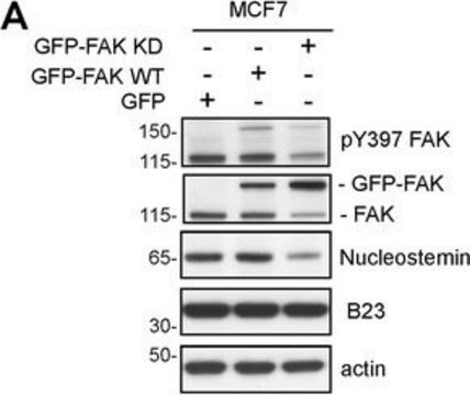
This Anti-phospho-Focal Adhesion Kinase (Tyr397) Antibody, clone 18 is validated for use in WB for the detection of phospho-Focal Adhesion Kinase (Tyr397).

Quality

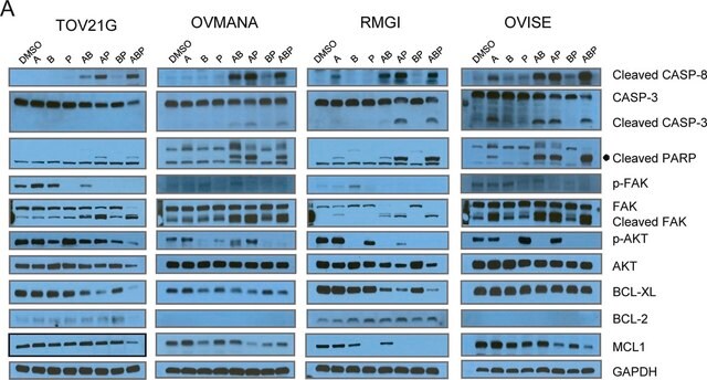
Evaluated by Western Blot in LPS treated RAW 264 lysates.
Western Blot Analysis: 1:500 dilution of this lot detected phospho-FAK (Tyr397) on 10 ug of LPS treated RAW 264 lysates.

Target description

125 kDa

Linkage

Replaces: 04-974

Physical form

Format: Purified
Protein A purified
Purified mouse monoclonal IgG1 in aqueous buffered solution containing 50% glycerol, BSA, and <0.09% sodium azide.

Storage and Stability

Stable for 1 year at -20ºC from date of receipt.
Note: Variability in freezer temperatures below -20°C may cause glycerol containing solutions to become frozen during storage.

Analysis Note

Control
LPS treated RAW 264 lysates.

Other Notes

Concentration: Please refer to the Certificate of Analysis for the lot-specific concentration.

Disclaimer

Unless otherwise stated in our catalog or other company documentation accompanying the product(s), our products are intended for research use only and are not to be used for any other purpose, which includes but is not limited to, unauthorized commercial uses, in vitro diagnostic uses, ex vivo or in vivo therapeutic uses or any type of consumption or application to humans or animals.

Not finding the right product?  

Try our Product Selector Tool.

Storage Class Code

10 - Combustible liquids

WGK

WGK 2

Flash Point(F)

Not applicable

Flash Point(C)

Not applicable


Certificates of Analysis (COA)

Search for Certificates of Analysis (COA) by entering the products Lot/Batch Number. Lot and Batch Numbers can be found on a product’s label following the words ‘Lot’ or ‘Batch’.

Already Own This Product?

Find documentation for the products that you have recently purchased in the Document Library.

Visit the Document Library

Melisa J Andrade et al.
Journal of cell science, 134(12) (2021-06-18)
UVBR-induced photolesions in genomic DNA of keratinocytes impair cellular functions and potentially determine the cell fate post-irradiation. The ability of insulin-like growth factor-I (IGF-I) to rescue epidermal keratinocytes after photodamage via apoptosis prevention and photolesion removal was recently demonstrated using
Catherine Moorwood et al.
Skeletal muscle, 4, 13-13 (2014-07-16)
The dystrophin glycoprotein complex (DGC) is located at the sarcolemma of muscle fibers, providing structural integrity. Mutations in and loss of DGC proteins cause a spectrum of muscular dystrophies. When only the sarcoglycan subcomplex is absent, muscles display severe myofiber
Derek L Clouthier et al.
Molecular and cellular biology, 35(9), 1573-1587 (2015-02-19)
Development of the cardiovascular system is critically dependent on the ability of endothelial cells (ECs) to reorganize their intracellular actin architecture to facilitate migration, adhesion, and morphogenesis. Nck family cytoskeletal adaptors function as key mediators of actin dynamics in numerous
Christoph Seiler et al.
PLoS biology, 10(9), e1001386-e1001386 (2012-09-14)
The signals that initiate cell invasion are not well understood, but there is increasing evidence that extracellular physical signals play an important role. Here we show that epithelial cell invasion in the intestine of zebrafish meltdown (mlt) mutants arises in
Ling Guo et al.
Nature communications, 10(1), 845-845 (2019-02-21)
Cell metabolism is strongly influenced by mechano-environment. We show here that a fraction of kindlin-2 localizes to mitochondria and interacts with pyrroline-5-carboxylate reductase 1 (PYCR1), a key enzyme for proline synthesis. Extracellular matrix (ECM) stiffening promotes kindlin-2 translocation into mitochondria

Our team of scientists has experience in all areas of research including Life Science, Material Science, Chemical Synthesis, Chromatography, Analytical and many others.

Contact Technical Service