05-916
Anti-P-Cadherin Antibody, clone 6A9
ascites fluid, clone 6A9, Upstate®
Sign Into View Organizational & Contract Pricing
All Photos(2)
About This Item
UNSPSC Code:
12352203
eCl@ss:
32160702
NACRES:
NA.43
Recommended Products
biological source
mouse
Quality Level
antibody form
ascites fluid
antibody product type
primary antibodies
clone
6A9, monoclonal
species reactivity
human
manufacturer/tradename
Upstate®
technique(s)
immunocytochemistry: suitable
immunoprecipitation (IP): suitable
western blot: suitable
NCBI accession no.
UniProt accession no.
shipped in
dry ice
target post-translational modification
unmodified
Gene Information
human ... CDH3(1001)
Specificity
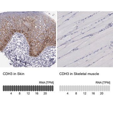
Recognizes P-Cadherin
Immunogen
P-Cadherin extracellular domain purified from A431 cell membranes.
Application
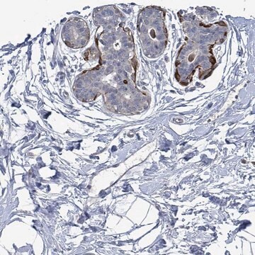
This Anti-P-Cadherin Antibody, clone 6A9 is validated for use in IP, WB, IC for the detection of P-Cadherin.
Quality
Routinely evaluated by immunoblot on A431 cell lysates.
Target description
Mr 120kDa
Analysis Note
Control
Positive Antigen Control: Catalog #12-301, non-stimulated A431 cell lysate. Add 2.5µL of 2-mercaptoethanol/100µL of lysate and boil for 5 minutes to reduce the preparation. Load 20µg of reduced lysate per lane for minigels.
Positive Antigen Control: Catalog #12-301, non-stimulated A431 cell lysate. Add 2.5µL of 2-mercaptoethanol/100µL of lysate and boil for 5 minutes to reduce the preparation. Load 20µg of reduced lysate per lane for minigels.
Legal Information
UPSTATE is a registered trademark of Merck KGaA, Darmstadt, Germany
Not finding the right product?
Try our Product Selector Tool.
Storage Class Code
10 - Combustible liquids
WGK
WGK 1
Certificates of Analysis (COA)
Search for Certificates of Analysis (COA) by entering the products Lot/Batch Number. Lot and Batch Numbers can be found on a product’s label following the words ‘Lot’ or ‘Batch’.
Already Own This Product?
Find documentation for the products that you have recently purchased in the Document Library.
K A Knudsen et al.
Human pathology, 31(8), 961-965 (2000-09-15)
Breast cancers often show reduced expression of the transmembrane cell-cell adhesion protein, E-cadherin. In addition, approximately half of breast carcinomas express P-cadherin, which correlates with poor survival. A large fragment of the E-cadherin extracellular domain can be detected in serum
M D Hines et al.
Journal of cell science, 112 ( Pt 24), 4569-4579 (1999-11-27)
Cadherin function is required for normal keratinocyte intercellular adhesion and stratification. In the present study, we have investigated whether cadherin-cadherin interactions may also modulate keratinocyte differentiation, as evidenced by alterations in the levels of several differentiation markers. Confluent keratinocyte cultures
Cadherin function is required for human keratinocytes to assemble desmosomes and stratify in response to calcium
Lewis, J. E., et al
The Journal of Investigative Dermatology, 102, 870-877 (1994)
M T Nieman et al.
Journal of cell science, 112 ( Pt 10), 1621-1632 (1999-04-23)
The cadherin/catenin complex mediates Ca2+-dependent cell-cell interactions that are essential for normal developmental processes. It has been proposed that sorting of cells during embryonic development is due, at least in part, to expression of different cadherin family members or to
Identification of human embryonic stem cell surface markers by combined membrane-polysome translation state array analysis and immunotranscriptional profiling.
Kolle G, Ho M, Zhou Q, Chy HS, Krishnan K, Cloonan N, Bertoncello I, Laslett AL, Grimmond SM
Stem Cells null
Our team of scientists has experience in all areas of research including Life Science, Material Science, Chemical Synthesis, Chromatography, Analytical and many others.
Contact Technical Service