Millicell® DCI Digital Cell Imager FAQs
Monitoring cell cultures is essential for assessing cell health and the viability of cell culture projects. The Millicell® DCI Digital Cell Imager helps researchers assess their cultures quickly and objectively, monitor cells, and measure cell confluency, cell count, and morphology. Here our team of scientists answers your frequently asked questions about the Millicell® DCI Digital Cell Imager.
Does the Millicell® DCI perform brightfield, phase, or fluorescence imaging? Can it capture time lapse movies and images?
Currently, the Millicell® DCI does not support true phase contrast or fluorescence imaging; the instrument can provide brightfield imaging and has some phase-like imaging capability. The Millicell® DCI is only able to capture static images, it does not support the capture of time lapse images and videos.
Currently the Millicell® DCI digital cell imager does not support remotely acquiring images, images must be taken in real time. Some image data can be processed and edited remotely once data is uploaded to the cloud. Mask and cell size can be adjusted in the cloud, however adjusting the image brightness must be done on the instrument when taking the image and cannot be edited in the cloud and images cannot be cropped or resized in the cloud as the scale bar and cell confluency estimates will be incorrect.
Images taken on the instrument are in PNG format and the exported data is in CSV format. The Millicell® DCI contains a camera with 5 MP CMOS sensor and resolution is 2560 x 1920, or monochrome and runs on the Dual Core IMX7 processor running at 1.2GHZ, so the image processing time is in milliseconds. The image area on the instrument for viewing is 2.4 mm x 1.4 mm.
How does the Millicell® DCI cell counting feature work?
The cell counting algorithm uses technology to determine area covered and finds the center of the individual areas. Millicell® DCI can also accurately count cells that do not have a nucleus, such as red blood cells, because it is not necessarily looking for a nucleus but rather the center of an individual spot.
For suspension cell lines, the cells need to fully settle onto the surface of the plate or be held in place, such as on a hemocytometer glass slide, before the Millicell® DCI can make an accurate cell count. The cell counting feature does not currently determine cell viability.
The Millicell® DCI can be used for cell monitoring and measuring cell confluency in any vessel that fits under the light source arm, including 6-well or 96-well plates. The LED light source is 2.2 inches away from the stage. The digital cell imager can also image in any vessel that fits under the light source arm but can only image a portion of a single well within a 96-well plate.
What magnification lens is on the Millicell® DCI?
The instrument has a 10X lens and is capable of magnification up to 20X using digital magnification.
Can I adjust the Millicell® DCI mask setting on the instrument or in the cloud?
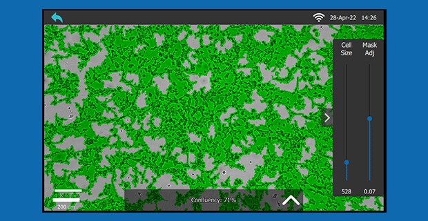
Mask settings can be changed on both the instrument and in the cloud.
From instrument:
After adjustment of mask in the cloud:


If I take several measurements without clicking “Begin Average”, can I average these measurements later in the cloud?
No, the “Begin Average” function needs to be used when taking the measurements to get an average for a single timepoint.
Can I access more information about a specific cell line from the cloud app?
Yes, you have the option to search our website for full information on different cell lines using the cell line specific feature.
Can I use all Millicell® DCI functions offline, and do I need a cloud account to save data to a USB drive?
Users can save images to a USB drive and use the device in offline mode even without a cloud account; approximately 10,000 images can be stored on the Millicell® DCI without cloud access. It is not critical to have a cloud account to use the instrument and all functions can be used offline. However, the user will not be able to manage, sort, or adjust parameters of images later.
When the device is used in offline mode any images that are taken cannot be uploaded to the cloud later. These images can be saved using a USB stick and used offline. If images are not uploaded at the time they were taken, the time stamp on the images will be when they upload to the cloud not the time they were taken.
If a group wants to buy multiple Millicell® DCI instruments for their various cell culture facilities, will all instruments share the same software subscription?
Yes, all the instruments can share the same software subscription, but the cloud account does not have unlimited storage space.
Is the Millicell® DCI compliant with 21 CFR Part 11 or GLP?
No, the digital cell imager is not 21 CFR Part 11 or Good Laboratory Practice (GLP) compliant.
How do I reset my PIN?
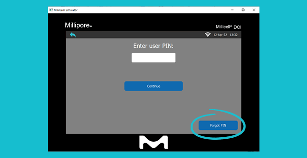
Users can remove their account by pressing on their name and tapping “Delete” and re-add their account. This will not hurt any of their data, they will just need to log in again. Users can also reset their PIN by pressing “Forgot PIN” (see image below). The system will ask for their email and password and allow them to reset the PIN.

Can the Millicell® DCI connect to a printer?
Currently the instrument cannot be connected to a printer. Images taken on the system can be downloaded from the cloud and printed from the computer.
Can the Millicell® DCI be used and stored in the cell culture hood?
Yes, Millicell® DCI is small enough to be used in the cell culture hood - the instrument measures 6 in by 11 in and weighs 6 lbs or 2.7kg. The instrument is stable from 5 – 40 oC and 20-80% relative humidity and is used between 15 – 30 oC or under general laboratory conditions.
Does the Millicell® DCI have an extended warranty?
The Millicell® DCI comes with a standard 1-year manufacturer warranty; currently there is no extended warranty.
Can the Millicell® DCI surface plate be heated?
The Millicell® DCI stage is not heated.
Are parameters displayed on the Millicell® DCI image?
All parameters of an image captured are displayed and can also be manipulated in the cloud web app. Cell masking, cell size and mask adjustment, and vessel size are examples of parameters that can be manipulated on the web app.
Pour continuer à lire, veuillez vous connecter à votre compte ou en créer un.
Vous n'avez pas de compte ?