Accéder au contenu
Merck
Toutes les photos(1)

Key Documents

236A-1

Sigma-Aldrich

CEA Rabbit Polyclonal Antibody

Se connecterpour consulter vos tarifs contractuels et ceux de votre entreprise/organisme


About This Item

Code UNSPSC :
12352203
Nomenclature NACRES :
NA.41

Source biologique

rabbit

Niveau de qualité

100
500

Conjugué

unconjugated

Forme d'anticorps

Ig fraction of antiserum

Type de produit anticorps

primary antibodies

Clone

polyclonal

Description

For In Vitro Diagnostic Use in Select Regions (See Chart)

Forme

buffered aqueous solution

Espèces réactives

human

Conditionnement

vial of 0.1 mL concentrate (236A-14)
vial of 0.5 mL concentrate (236A-15)
bottle of 1.0 mL predilute (236A-17)
vial of 1.0 mL concentrate (236A-16)
bottle of 7.0 mL predilute (236A-18)

Fabricant/nom de marque

Cell Marque

Technique(s)

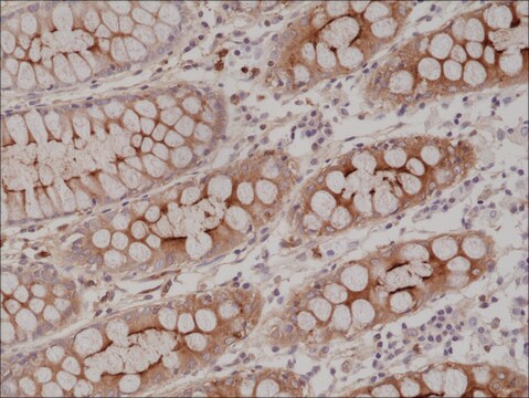
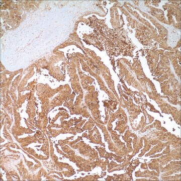
immunohistochemistry (formalin-fixed, paraffin-embedded sections): 1:200-1:500

Contrôle

colon

Conditions d'expédition

wet ice

Température de stockage

2-8°C

Visualisation

cytoplasmic

Informations sur le gène

human ... CEACAM5(1048)

Description générale

Anti-CEA is employed essentially as a tool to assist in the distinction between adenocarcinoma and epithelioid malignant mesotheliomas, along with other markers such as those against calretinin, CK 5&6, CD15, HBME-1, MOC-31, and Ber-EP4. Another suggested use of anti-CEA is to immunophenotype various metastatic adenocarcinomas as a means of identifying their origin within a panel of different markers. Anti-CEA positivity is seen in adenocarcinomas from the lung, colon, stomach, esophagus, pancreas, gallbadder, urachus, salivary gland, ovary, and endocervix. Polyclonal anti-CEA is useful in staining hepatocellular carcinoma in a canalicular pattern.

Liaison

CEA Positive Control Slides, Product No. 236S, are available for immunohistochemistry (formalin-fixed, paraffin-embedded sections).

Forme physique

Solution in Tris Buffer, pH 7.3-7.7, with 1% BSA and <0.1% Sodium Azide

Notes préparatoires

Download the IFU specific to your product lot and formatNote: This requires a keycode which can be found on your packaging or product label.

Autres remarques

For Technical Service please contact: 800-665-7284 or email: service@cellmarque.com

Informations légales

Cell Marque is a trademark of Merck KGaA, Darmstadt, Germany

Vous ne trouvez pas le bon produit ?  

Essayez notre Outil de sélection de produits.


Certificats d'analyse (COA)

Recherchez un Certificats d'analyse (COA) en saisissant le numéro de lot du produit. Les numéros de lot figurent sur l'étiquette du produit après les mots "Lot" ou "Batch".

Déjà en possession de ce produit ?

Retrouvez la documentation relative aux produits que vous avez récemment achetés dans la Bibliothèque de documents.

Consulter la Bibliothèque de documents

A Alkushi et al.
Virchows Archiv : an international journal of pathology, 442(3), 271-277 (2003-03-21)
Adenocarcinomas of the uterine cervix show a wide range of morphological features, and can be confused with endometrial adenocarcinoma in biopsy or curetting specimens. The objective of this study was to use tissue microarray technology to evaluate the immunoprofile of
Carl Morrison et al.
Modern pathology : an official journal of the United States and Canadian Academy of Pathology, Inc, 15(12), 1279-1287 (2002-12-14)
The distinction of hepatocellular carcinoma (HCC) from metastatic adenocarcinoma (MA) and cholangiocarcinoma (CC) in some cases requires the use of immunohistochemistry. CD10 has recently been suggested as a useful stain for HCC. We directly compared CD10 with other immunohistochemical markers
K Sheahan et al.
American journal of clinical pathology, 94(2), 157-164 (1990-08-01)
To evaluate the role of carcinoembryonic antigen (CEA) in solving problems of tumor histogenesis in surgical pathology, monoclonal antibodies to four distinct epitopes of CEA (E-Z-EM) were applied to paraffin sections of 303 epithelial neoplasms from multiple sites. Two epitopes
P W Shield et al.
American journal of clinical pathology, 105(2), 157-162 (1996-02-01)
The contribution of immunocytochemical (ICC) staining to the diagnosis of a range of cytologic specimens reported over a 20-month period was retrospectively assessed. A total of 194 cases (1.6% of total workload) were stained for diagnostic purposes in this period.

Notre équipe de scientifiques dispose d'une expérience dans tous les secteurs de la recherche, notamment en sciences de la vie, science des matériaux, synthèse chimique, chromatographie, analyse et dans de nombreux autres domaines..

Contacter notre Service technique