Skip to Content
Merck
All Photos(3)

Key Documents

06-498-I

Sigma-Aldrich

Anti-PDGFR Beta Antibody

from rabbit, purified by affinity chromatography

Synonym(s):

Platelet-derived growth factor receptor beta, Beta platelet-derived growth factor receptor, Beta-type platelet-derived growth factor receptor, CD140 antigen-like family member B, CD140b, PDGF-R-beta, PDGFR-1, PDGFR-beta, Platelet-derived growth factor re

Sign Into View Organizational & Contract Pricing


About This Item

UNSPSC Code:
12352203
eCl@ss:
32160702

biological source

rabbit

Quality Level

antibody form

affinity isolated antibody

antibody product type

primary antibodies

clone

polyclonal

purified by

affinity chromatography

species reactivity

mouse

species reactivity (predicted by homology)

human (based on 100% sequence homology), bovine (based on 100% sequence homology), canine (based on 100% sequence homology)

technique(s)

immunocytochemistry: suitable
immunoprecipitation (IP): suitable
western blot: suitable

NCBI accession no.

UniProt accession no.

shipped in

ambient

target post-translational modification

unmodified

Gene Information

human ... PDGFRB(5159)

General description

Platelet-derived growth factor receptor beta (EC 2.7.10.1; UniProt P09619; also known as Beta platelet-derived growth factor receptor, Beta-type platelet-derived growth factor receptor, CD140 antigen-like family member B, CD140b, PDGF-R-beta, PDGFR-1, PDGFR-beta, Platelet-derived growth factor receptor 1) is encoded by the PDGFRB (also known as IBGC4, IMF1, PDGFR, PDGFR1) gene (Gene ID 5159) in human. PDGFR-1 is a tyrosine-protein kinase that acts as a cell-surface receptor for homodimeric PDGFB and PDGFD and for heterodimers formed by PDGFA and PDGFB. PDGF receptors consist of extracellular domains with five immunoglobulin (Ig) - like domains and intracellular parts with kinase domains, which contain characteristic inserts of about 100 amino acid residues without homology to kinases. Ligand binding occurs mainly to Ig-like domains 2 and 3, which causes dimerization of the receptor, which is further stabilized by direct receptor-receptor interactions involving Ig-like domain 4. In the absence of a bound ligand, receptor is present in an inactive conformation. However, upon ligand binding it undergoes dimerization and autophosphorylation on tyrosine residues. PDGFR-1m plays an essential role in the regulation of embryonic development, cell proliferation, survival, differentiation, chemotaxis, and migration. It is also considered to be essential for endothelial cell proliferation, migration and recruitment of pericytes and smooth muscle cells to form blood vessels. PDGFR-1 phosphorylates phospholipase C-gamma 1 (PLCG1), PIK3R1, PTPN11, RASA1/GAP, CBL, SHC1 and NCK1. Activation of PLCG1 leads to the production of diacylglycerol and IP3 that are involved in calcium mobilization and activation of PKC. Ref.: Heldin C-H (2013). Cell Commun. Signaling 11:97.C1

Specificity

This polyclonal antibody targets a cytoplasmic sequence C-terminal to the PDGFR kinase domain.

Immunogen

KLH-conjugated linear peptide corresponding to a cytoplasmic sequence C-terminal to the kinase domain of human PDGFR .

Application

Detect Platelet-derived growth factor receptor beta using this rabbit polyclonal Anti-PDGFR Beta Antibody, Cat. No. 06-498-I, validated for use in Immunocytochemistry, Immunoprecipitation, and Western Blotting.
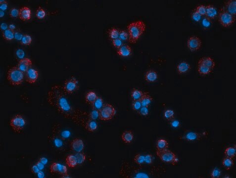
Immunocytochemistry Analysis: A 1:100 dilution from a representative lot detected PDGFR immunoreactivity in 4% paraformaldehyde-fixed, 0.3% Triton X-100-permeabilized BALB/3T3 clone A31 cells.

Immunoprecipitation Analysis: 5 µg from a representative lot immunoprecipitated PDGFR from 500 µg of BALB/3T3 clone A31 cell lysate.
Research Category
Signaling

Quality

Evaluated by Western Blotting in BALB/3T3 clone A31 cell lysate.

Western Blotting Analysis: 2 µg/mL of this antibody detected PDGFR in 10 µg of BALB/3T3 clone A31 cell lysate.

Target description

~180 kDa observed. Target band size appears larger than the calculated molecular weight of 120.6/119.6 kDa (human/mouse; signal peptide removed) due to glycosylation. Uncharacterized bands may be observed in some lysate(s).

Linkage

Replaces: 06-498

Physical form

Affinity purified.
Purified rabbit polyclonal antibody in buffer containing 0.1 M Tris-Glycine (pH 7.4), 150 mM NaCl with 0.05% sodium azide.

Storage and Stability

Stable for 1 year at 2-8°C from date of receipt.

Other Notes

Concentration: Please refer to lot specific datasheet.

Disclaimer

Unless otherwise stated in our catalog or other company documentation accompanying the product(s), our products are intended for research use only and are not to be used for any other purpose, which includes but is not limited to, unauthorized commercial uses, in vitro diagnostic uses, ex vivo or in vivo therapeutic uses or any type of consumption or application to humans or animals.

Not finding the right product?  

Try our Product Selector Tool.

Storage Class Code

12 - Non Combustible Liquids

WGK

WGK 1

Flash Point(F)

Not applicable

Flash Point(C)

Not applicable


Certificates of Analysis (COA)

Search for Certificates of Analysis (COA) by entering the products Lot/Batch Number. Lot and Batch Numbers can be found on a product’s label following the words ‘Lot’ or ‘Batch’.

Already Own This Product?

Find documentation for the products that you have recently purchased in the Document Library.

Visit the Document Library

María Valero-Muñoz et al.
Circulation. Heart failure, 14(9), e008372-e008372 (2021-08-20)
The management of clinical heart failure with a preserved ejection fraction (HFpEF) is often complicated by concurrent renal dysfunction, known as the cardiorenal syndrome. This, combined with the notable lack of evidence-based therapies for HFpEF, highlights the importance of examining

Our team of scientists has experience in all areas of research including Life Science, Material Science, Chemical Synthesis, Chromatography, Analytical and many others.

Contact Technical Service