Recommended Products
manufacturer/tradename
Chemicon®
technique(s)
ELISA: suitable
UniProt accession no.
Gene Information
General description
Vascular cell adhesion molecule-1 (VCAM-1, CD106) is a member of the immunoglobulin gene superfamily. Two forms of VCAM-1 with six or seven extracellular Ig-like domains arise due to alternative splicing from a seven-domain VCAM-1 (7D VCAM-1). 7D VCAM-1 is the dominant form expressed by cultured human endothelial cells [Cybulsky et al., 1991; Hession et al., 1991]. Homology of domains 1 through 3 with domains 4 through 6 suggests a gene duplication event [Cybulsky et al., 1991; Hession et al., 1991]. The cDNA of 7D VCAM-1 predicts a core protein of approximately 81 kDa with seven sites for N-linked glycosylation. Upon complete glycosylation the mature protein has a molecular weight of approximately 102 kDa. This observation is in general agreement with the 110 kDa protein immunoprecipitated from cytokine-activated endothelium [Carlos et al., 1990; Rice et al., 1990].
VCAM-1 appears to have been highly conserved through evolution. Both rat and mouse VCAM-1 are highly homologous at the protein level to the human VCAM-1 (77% and 76%, respectively) [Hession et al., 1992].
VCAM-1 supports the adhesion of lymphocytes, monocytes, natural killer cells, eosinophils and basophils, through its interaction with integrin a4b1 (VLA-4). VCAM-1/VLA-4 interaction mediates firm adherence of circulating non-neutrophilic leukocytes to endothelium [Sharar et al., 1995]. VCAM-1 also participates in leukocyte adhesion outside of the vasculature, mediating precursor lymphocyte adhesion to bone marrow stromal cells [Ryan et al. 1991] and B cell binding to lymph node follicular dendritic cells [Feedman et al., 1992]. VCAM-1 is not constitutively expressed on endothelium, but can be up-regulated in vitro in response to LPS, TNF-a, and IL-1 [Carlos & Harlan, 1990; Osborn et al., 1989], as well as to interferon-g and IL-4 [Masinovsky et al. 1990]. VCAM-1 is also present on tissue macrophages, dendrite cells, bone marrow fibroblasts, myoblasts and myotubes.
A soluble form of VCAM-1 has been described [De Maeyer & De Maeyer-Buignard, 1995; Lobb et al., 1991], and soluble VCAM-1 has been found in serum [Gearing et al., 1995]. Increased levels of soluble VCAM-1 can be detected in several disease states, including cancer [Banks et al., 1993; Fortis et al., 1995; Gearing & Newman, 1993; Lai et al., 1995], autoimmune disorders [Gearing & Newman, 1993; Doreduffy et al., 1995; Gruschwitz et al., 1995; Mason et al., 1993; Mrowka & Sieberth, 1995; Spronk et al., 1994; Wellicome et al., 1993], infections [Jakobsen et al., 1994] and inflammations [Gearing & Newman, 1993; Mrowka & Sieberth, 1995; Adams et al., 1995; Lim et al., 1995].
Test Principle:
An anti-VCAM-1 monoclonal antibody is adsorbed onto microwells.
Soluble VCAM-1 present in a sample or standard then binds to antibodies adsorbed to the microwells. A mixture of biotin-conjugated monoclonal anti-VCAM-1 antibody and Streptavidin-HRP conjugate is added. Biotinylated anti-VCAM-1 antibody binds to VCAM-1 captured by the first antibody.
Streptavidin-HRP binds to the biotinylated anti-VCAM-1. Unbound biotinylated anti-VCAM-1 and Streptavidin-HRP conjugate is removed with a wash step and HRP substrate solution is added to the wells.
The pair of monoclonal antibodies used in this ELISA detect the soluble form of VCAM-1 present in serum, plasma, urine, and other biological fluids.
An amount of colored product is formed, proportional to the amount of soluble VCAM-1 present in the sample. The reaction is terminated by addition of acid and absorbance is measured at 450 nm. A standard curve is prepared from six soluble VCAM-1 standard dilutions and the VCAM-1 sample concentration is determined.
Specificity
Analytes Available: sVCAM-1
Detection Methods: Chromogenic
Detection Methods: Chromogenic
Application
This Human sVCAM-1 ELISA is used to measure & quantify sVCAM-1 levels in Inflammation & Immunology & Neuroscience research.
Features and Benefits
- 5 mL and 10 mL graduated pipettes
- 5 μL to 1000 μL adjustable single channel micropipettes with disposable tips
- 50 μL to 300 μL adjustable multichannel micropipette with disposable tips
- Multichannel micropipette reservoir - Beakers, flasks, cylinders necessary for preparation of reagents
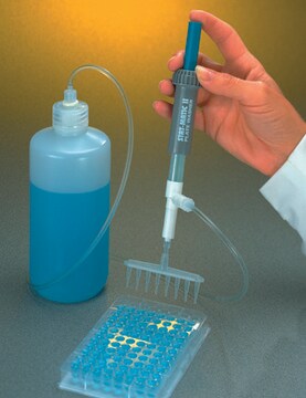
- Device for delivery of wash solution (multichannel wash bottle or automatic wash system)
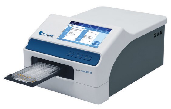
- Microwell strip reader capable of reading at 450 nm (620 nm as optional reference wavelength)
- Glass-distilled or deionized water - Statistical calculator with program to perform linear regression analysis.
Components
- 2 aluminum pouches with 3 Microwell Strips (16 wells per strip) coated with Monoclonal Antibody (murine) to human VCAM-1
- 1 vial (0.1 mL) Conjugate Mixture containing Biotin-Conjugated Anti-VCAM-1 Monoclonal (murine) Antibody mixed with Streptavidin-HRP*
- 2 vials (200 ng/mL as reconstituted) soluble VCAM-1 Standard, lyophilized*
- 1 vial lyophilized control serum· 1 bottle (50 mL) Wash Buffer Concentrate 20X (PBS with 1% Tween 20)*
- 1 vial (5 mL) Assay Buffer Concentrate 20X (PBS with 1% Tween 20 and 10% BSA)*
- 1 vial (7 mL) Substrate Solution I (tetramethyl-benzidine)
- 1 vial (7 mL) Substrate Solution II (0.02 % buffered hydrogen peroxide)
- 1 vial (12 mL) Stop Solution (1M Phosphoric Acid, H3PO4), ready to use
- 1 Microwell Strip Holder· 2 Adhesive Plate Covers
- Reagents containing 0.01% thimerosal as preservative
Storage and Stability
Precautions:
- Reagents are intended for research use only and are not for use in diagnostic or therapeutic procedures.
- Do not mix or substitute reagents with those from other lots or other sources.
- Do not use kit reagents beyond expiration date on label.
- Do not expose kit reagents to strong light during storage or incubation.
- Rubber or disposable latex gloves should be worn while handling kit reagents or specimens.
- Some reagents contain thimerosal as preservative, which is highly toxic by inhalation, ingestion, or contact with skin. Thimerosal is a possible mutagen and should be handled accordingly.
- Avoid contact of substrate solutions with oxidizing agents and metal.
- In order to avoid microbial contamination or cross-contamination of reagents or specimens which may invalidate the test use disposable pipette tips and/or pipettes.
- Use clean, dedicated reagent trays for dispensing the conjugate and substrate reagents.
- Exposure to acids will inactivate the conjugate.
- Glass-distilled water or deionized water must be used for reagent preparation.
- Substrate solutions must be at room temperature prior to use.
- Since exact conditions may vary from assay to assay, a standard curve must be established for every run.
- Bacterial or fungal contamination of either screen samples or reagents or cross-contamination between reagents may cause erroneous results.
- Disposable pipette tips, flasks or glassware are preferred. Reusable glassware must be washed and thoroughly rinsed of all detergents before use.
- Improper or insufficient washing at any stage of the procedure will result in either false positive or false negative results. Completely empty wells before dispensing fresh Wash Buffer, fill with Wash Buffer as indicated for each wash cycle and do not allow wells to sit uncovered or dry for extended periods.
- Any human anti-mouse IgG antibody (HAMA) present may interfere with assays utilizing murine monoclonal antibodies leading to both false positive and false negative results. HAMA interference can be reduced by adding murine immunoglobulins (serum, ascitic fluid, or monoclonal antibodies of irrelevant specificity) to the Sample Diluent.
Legal Information
CHEMICON is a registered trademark of Merck KGaA, Darmstadt, Germany
Disclaimer
Unless otherwise stated in our catalog or other company documentation accompanying the product(s), our products are intended for research use only and are not to be used for any other purpose, which includes but is not limited to, unauthorized commercial uses, in vitro diagnostic uses, ex vivo or in vivo therapeutic uses or any type of consumption or application to humans or animals.
Certificates of Analysis (COA)
Search for Certificates of Analysis (COA) by entering the products Lot/Batch Number. Lot and Batch Numbers can be found on a product’s label following the words ‘Lot’ or ‘Batch’.
Already Own This Product?
Find documentation for the products that you have recently purchased in the Document Library.
Our team of scientists has experience in all areas of research including Life Science, Material Science, Chemical Synthesis, Chromatography, Analytical and many others.
Contact Technical Service
