03-206
RIPAb+ hnRNP U - RIP Validated Antibody and Primer Set
clone 3G6, from mouse
Sinonimo/i:
Scaffold attachment factor A, heterogeneous nuclear ribonucleoprotein U, heterogeneous nuclear ribonucleoprotein U (scaffold attachment factor A), hnRNP U, hnRNP U protein, p120 nuclear protein
About This Item
Prodotti consigliati
Origine biologica
mouse
Livello qualitativo
Forma dell’anticorpo
purified immunoglobulin
Clone
3G6, monoclonal
Reattività contro le specie
human
Produttore/marchio commerciale
RIPAb+
Upstate®
tecniche
RIP: suitable
western blot: suitable
Isotipo
IgG2bκ
N° accesso NCBI
N° accesso UniProt
Condizioni di spedizione
dry ice
Descrizione generale
Proteins of the heterogeneous nuclear ribonucleoparticles (hnRNP) family form a structurally diverse group of RNA binding proteins implicated in various functions. Recently, hnRNP proteins have been shown to hinder communication between factors bound to different splice sites. hnRNP-U, also termed scaffold attachment factor A (SAF-A), binds to pre-mRNA and nuclear matrix/scaffold attachment region DNA elements.
Specificità
Immunogeno
Applicazioni
Representative lot data.
RIP lysate from HeLa cells (~2 X 10E7 cell equivalents per IP) was subjected to immunoprecipitation using 5 µg of either a normal mouse IgG, (Cat. # CS200621), or 5 µg of Anti-hnRNP U antibody (Cat. # CS207320). ten percent of the precipitated proteins (lane 1: normal mouse IgG, lane 2: hnRNP U) and HeLa whole cell lysate (lane 3) were resolved by electrophoresis, transferred to nitrocellulose and probed with anti-hnRNP U antibody (Cat. # CS207320, 1:1000). Proteins were visualized using One-Step IP-Western kit (GenScript Cat. # L00231).
Arrow indicates hnRNP U. (Figure 2).
Automated Microfluidics-based Electrophoretic RNA Separation and Analysis (MFE):
Representative lot data.
RIP Lysate prepared from HeLa cells (2 X 10E7 cell equivalents per IP) were subjected to immunoprecipitation using 5 µg of either 1. normal mouse IgG (Cat. # CS200621), or 2. Anti-hnRNP U antibody (Cat. # CS207320) and the Magna RIP RNA-Binding Protein Immunoprecipitation Kit (Cat. # 17-700).
Successful immunoprecipitation of hnRNP U-associated RNA was verified by automated microfluidics-based electrophoretic RNA separation and analysis. Please refer to the Magna RIP (Cat. # 17-700) or EZ-Magna RIP (Cat. # 17-701) protocol for experimental details. Electropherograms were generated by plotting fluorescence intensities versus migration times (Figure 3A). The virtual gel view was created from this data (Figure 3B).
Western Blot Analysis:
Representative lot data.
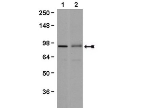
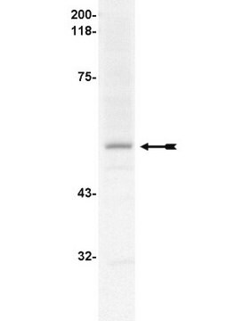
K562 cell lysate was probed with Anti-hnRNP U, clone 3G6 (0.01 µg/mL). Proteins were visualized using a Goat Anti-Mouse IgG secondary antibody conjugated to HRP and a chemiluminescence detection system.
Arrow indicates hnRNP U (~120 kDa). (Figure 4).
Epigenetics & Nuclear Function
RNA Metabolism & Binding Proteins
Apoptosis - Additional
Confezionamento
Qualità
Representative lot data.
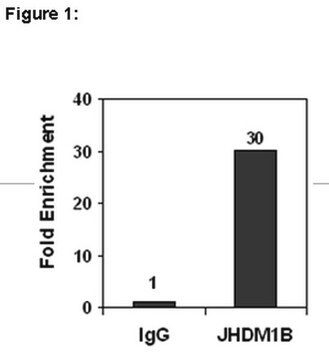
RIP Lysate prepared from HeLa cells (2 X 10E7 cell equivalents per IP) were subjected to immunoprecipitation using 5 µg of either a normal mouse IgG or 5 µg of Anti-hnRNP U antibody and the Magna RIP® RNA-Binding Protein Immunoprecipitation Kit (Cat. # 17-700).
Successful immunoprecipitation of hnRNP U-associated RNA was verified by qPCR using RIP Primers Ribosomal Protein S19, (Figure 1).
Please refer to the Magna RIP (Cat. # 17-700) or EZ-Magna RIP (Cat. # 17-701) protocol for experimental details.
Descrizione del bersaglio
Stato fisico
Normal Mouse IgG, Part # CS200621. One vial containing 125 µg of purified mouse IgG in 125 µL of storage buffer containing 0.1% sodium azide. Store at -20°C.
RIP Primers, Ribosomal Protein S19, Part # CS207321. One vial containing 75 μL of 5 μM of each primer specific for human c-myc 3′ UTR. Store at -20°C.
FOR: ACG CGA GCT GCT TCC ACA G
REV: AGC TGC CAC CTG TCC GGC
Stoccaggio e stabilità
Handling Recommendations: Upon receipt, and prior to removing the cap, centrifuge the vial and gently mix the solution. Aliquot into microcentrifuge tubes and store at -20°C. Avoid repeated freeze/thaw cycles, which may damage IgG and affect product performance. Note: Variabillity in freezer temperatures below -20°C may cause glycerol containing solutions to become frozen during storage.
Risultati analitici
Includes negative control normal mouse IgG antibody and control primers specific for the cDNA of human Ribosomal Protein S19.
Altre note
Note legali
Esclusione di responsabilità
Codice della classe di stoccaggio
10 - Combustible liquids
Certificati d'analisi (COA)
Cerca il Certificati d'analisi (COA) digitando il numero di lotto/batch corrispondente. I numeri di lotto o di batch sono stampati sull'etichetta dei prodotti dopo la parola ‘Lotto’ o ‘Batch’.
Possiedi già questo prodotto?
I documenti relativi ai prodotti acquistati recentemente sono disponibili nell’Archivio dei documenti.
Il team dei nostri ricercatori vanta grande esperienza in tutte le aree della ricerca quali Life Science, scienza dei materiali, sintesi chimica, cromatografia, discipline analitiche, ecc..
Contatta l'Assistenza Tecnica.