Accéder au contenu
MilliporeSigma
Toutes les photos(1)

Documents

R5403

Sigma-Aldrich

Monoclonal Anti-Rhodopsin antibody produced in mouse

clone 1D4, purified immunoglobulin, buffered aqueous solution

Synonyme(s) :

Anti-CSNBAD1, Anti-OPN2, Anti-RP4

Se connecterpour consulter vos tarifs contractuels et ceux de votre entreprise/organisme


About This Item

Numéro MDL:
Code UNSPSC :
12352203
Nomenclature NACRES :
NA.41

Source biologique

mouse

Niveau de qualité

Conjugué

unconjugated

Forme d'anticorps

purified immunoglobulin

Type de produit anticorps

primary antibodies

Clone

1D4, monoclonal

Forme

buffered aqueous solution

Espèces réactives

human, rat, bovine

Technique(s)

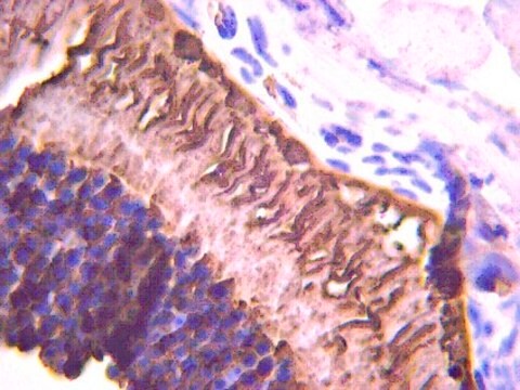
immunocytochemistry: 1:1,000 using human retinal samples
western blot: 1:1,000 using Sf9 cells expressing the bovine gene

Isotype

IgG1

Numéro d'accès UniProt

Conditions d'expédition

wet ice

Température de stockage

−20°C

Modification post-traductionnelle de la cible

unmodified

Informations sur le gène

human ... RHO(6010)
rat ... Rho(24717)

Description générale

Vision involves the conversion of light into electrochemical signals that are processed by the retina and subsequently sent to and interpreted by the brain. The process of converting light to an electrochemical signal begins when the membrane-bound protein, rhodopsin, absorbs light within the retina. Photoexcitation of rhodopsin causes the cytoplasmic surface of the protein to become catalytically active. In the active state, rhodopsin activates transducin, a GTP binding protein. Once activated, transducin promotes the hydrolysis of cGMP by phosphodiesterase (PDE). The decrease of intracellular cGMP concentrations causes the ion channels within the outer segment of the rod or cone to close, thus causing membrane hyperpolarization and, eventually, signal transmission. Rhodopsin′s activity is believed to be shut off by its phosphorylation followed by binding of the soluble protein arrestin.

Spécificité

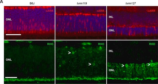
By Western blot, mouse monoclonal clone 1D4 anti-Rhodopsin antibody detects an ~40 kDa protein representing rhodopsin from Sf 9 cells expressing the bovine gene. Immunocytochemical staining of rhodopsin in human retinal samples results in staining of both rod and cone outer segments.
The epitope for this antibody has been localized to the C-terminal nine amino acids of bovine rhodopsin known as the 1D4 epitope, and detects rhodopsin from human and bovine retinal samples.

Immunogène

bovine rhodopsin.

Application

Applications in which this antibody has been used successfully, and the associated peer-reviewed papers, are given below.
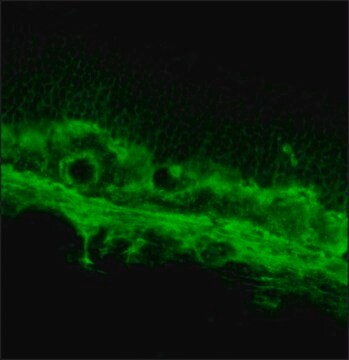
Immunohistochemistry (1 paper)
Mouse monoclonal clone 1D4 anti-Rhodopsin antibody has been successfully used in Western blot, immunocytochemistry, and immunoprecipitation. It is used as a probe to determine the presence and roles of rhodopsin in the conversion of light into electrochemical signals.

Forme physique

Solution in phosphate buffered saline containing 1 mg/mL BSA and 0.05% sodium azide.

Clause de non-responsabilité

Unless otherwise stated in our catalog or other company documentation accompanying the product(s), our products are intended for research use only and are not to be used for any other purpose, which includes but is not limited to, unauthorized commercial uses, in vitro diagnostic uses, ex vivo or in vivo therapeutic uses or any type of consumption or application to humans or animals.

Not finding the right product?  

Try our Outil de sélection de produits.

Code de la classe de stockage

10 - Combustible liquids

Classe de danger pour l'eau (WGK)

nwg

Point d'éclair (°F)

Not applicable

Point d'éclair (°C)

Not applicable


Certificats d'analyse (COA)

Recherchez un Certificats d'analyse (COA) en saisissant le numéro de lot du produit. Les numéros de lot figurent sur l'étiquette du produit après les mots "Lot" ou "Batch".

Déjà en possession de ce produit ?

Retrouvez la documentation relative aux produits que vous avez récemment achetés dans la Bibliothèque de documents.

Consulter la Bibliothèque de documents

Vadim Y Arshavsky
Trends in neurosciences, 25(3), 124-126 (2002-02-20)
Rhodopsin phosphorylation has provided one of the first examples of the ubiquitous regulatory pattern of specific kinases downregulating the activity of G-protein-coupled receptors. However, only recently have studies in living animals allowed us to consider the role of rhodopsin phosphorylation
Maria Carmela Latella et al.
Molecular therapy. Nucleic acids, 5(11), e389-e389 (2016-11-23)
The bacterial CRISPR/Cas system has proven to be an efficient tool for genetic manipulation in various organisms. Here we show the application of CRISPR-Cas9 technology to edit the human Rhodopsin (RHO) gene in a mouse model for autosomal dominant Retinitis
Transduction mechanisms of vertebrate and invertebrate photoreceptors.
S Yarfitz et al.
The Journal of biological chemistry, 269(20), 14329-14332 (1994-05-20)
Jun Yin et al.
Investigative ophthalmology & visual science, 53(8), 4943-4951 (2012-06-30)
In experimental eye research, zebrafish has become a powerful model for human retina disorders. The purpose of the present study is the characterization of antibodies commonly employed in zebrafish models for rod photoreceptor degeneration. The 1D4 monoclonal antibody, developed against
Min Zou et al.
Developmental dynamics : an official publication of the American Association of Anatomists, 244(1), 86-97 (2014-09-03)
During retinal and spinal cord neurogenesis, Notch signaling plays crucial roles in regulating proliferation and differentiation of progenitor cells. One of the Notch ligands, Delta-like 4 (Dll4), has been shown to be expressed in subsets of retinal and spinal cord

Notre équipe de scientifiques dispose d'une expérience dans tous les secteurs de la recherche, notamment en sciences de la vie, science des matériaux, synthèse chimique, chromatographie, analyse et dans de nombreux autres domaines..

Contacter notre Service technique