Validated Cell Types for Millicell® DCI Digital Cell Imager
The Millicell® DCI Digital Cell Imager enables quick, objective measurement of common cell culture parameters including confluency, cell count, and morphology across an extensive range of adherent cell cultures. Measurements can be made directly in the flask, plate, or vessel, or using a hemocytometer. These objective results, combined with growth trend analysis, allow for more consistent cell cultures. Our R&D scientists have been working to validate the Millicell® DCI on some of the most common and trickiest cells. Select a cell line or cell type to view the data.
A431 Cell Line
- Catalog number: 85090402
- Source: Human skin (female), squamous carcinoma
- Cell type/morphology: Adherent, epithelial
- Application: To study p38 MAPK-induced apoptosis, to study melanoma-specific CD44 receptor, to study the effect of anti-cancer medical plants
- Biosafety level: 2
- Handling: Easy
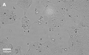
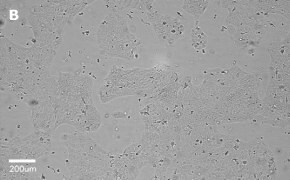
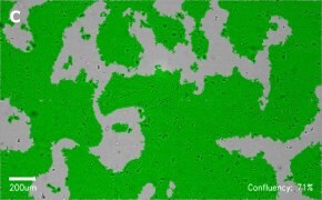
- Images: Brightfield image on Millicell® DCI at 10X (A), 20X (B) and the 10X image with confluency mask (C).
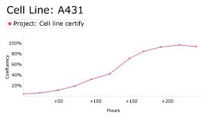
8. Growth Curve
9. Cell culture media:
EMEM (EBSS) + 2 mM L-Glutamine (G7513) + 1% Non-Essential Amino Acids (NEAA) + 10% FBS (F2442).
Split sub-confluent cultures (70-80%) 1:3 to 1:6, i.e., seeding at 2-4 x 10,0000 cells/cm2 using 0.25% trypsin or trypsin/EDTA; 5% CO2; 37 °C.
10. Protocols: Cell culture protocols are available in product datasheet.
A549 Cell Line
- Catalog number: 86012804
- Source: Human, lung (carcinoma)
- Cell type/morphology: Adherent, epithelial
- Application: To study the role of BChTT enzyme in cancer inhibition, to study the effects of insulin and IGF1, to study alveolar type II differentiation
- Biosafety level: 2
- Handling: Easy
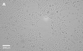
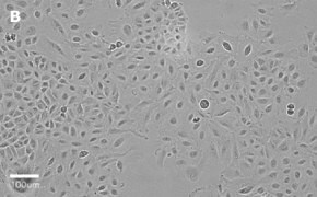
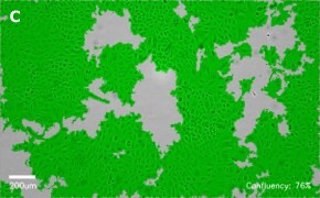
- Images: Brightfield image on Millicell® DCI at 10X (A), 20X (B) and the 10X image with confluency mask (C).
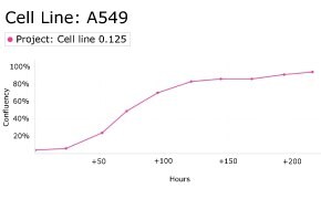
8. Growth Curve
ALMC-1 Cell Line
- Catalog number: SCC430
- Source: Human bone marrow
- Cell type/morphology: Suspension cell line
- Application: Plasma cell myeloma/amyloidosis
- Biosafety level: 2
- Handling: Medium
- Images: Brightfield image on Millicell® DCI (A) and flow cytometry histogram showing expression of CD44 in yellow and unstained cells in gray (B)


- Genetic profile:
- Cell culture media:
Cells were thawed in ALMC-1 recovery medium and expanded in ALMC-1 Expansion Medium comprised of IMDM medium (I6529) with 10% FBS (ES-009-B), 2 mM L-Glutamine (TMS-002-C), 2 ng/mL IL-6 (GF338) and 1X penicillin/streptomycin (TMS-AB2-C, optional). - Protocols: Cell culture protocols are available in product datasheet.
- Scepter™ histogram: Cells were counted on a Scepter™ 3.0 automated cell counter.

Caco-2 Cell Line
- Catalog number: 86010202
- Source: Human colon (Caucasian colon adenocarcinoma)
- Cell type/morphology: Adherent, epithelial
- Application: To study if components of the sigma B regulon in Listeria monocytogenes contribute to cell invasion, for functional analysis of bacteriocin divercin AS7, to study the intracellular invasion by Listeria ivanovii
- Biosafety level: 2
- Handling: Easy
- Images: Brightfield image on Millicell® DCI at 10X (A), 20X (B), and the 10X image with confluency mask (C).


- Growth curve:

- Cell culture media:
EMEM (EBSS) (M2279) + 2 mM Glutamine (G7513) + 1% Non-Essential Amino Acids (NEAA) (M7145) + 10% Fetal Bovine Serum (FBS) (ES-009-B).
Split sub-confluent cultures (70-80%) 1:3 to 1:6, i.e. seeding at 2-4 x 10,000 cells/cm2 using 0.25% Trypsin or Trypsin/EDTA; 5% CO2; 37°C. During routine subculture, the cells should always be subcultured before they achieve confluence. Cells may show the appearance of circular vacuoles in the cytoplasm. These may increase in frequency as the culture density increases to confluence. To reduce their frequency, media change confluent cultures after 2-3 days if not subcultured. Cells can clump if not separated into a single-cell suspension when split. - Protocols: Cell culture protocols are available in the product datasheet.
- Scepter™ histogram: Cells were counted on a Scepter™ 3.0 automated cell counter.

CHO-K1 Cell Line
- Catalog number: 85051005
- Source: Non-human; hamster ovary
- Cell type/morphology: Adherent, epithelial
- Application: Nutritional and gene expression studies have been used to study the effect of plasmid pCM01 induction on the association of bacteria with CHO-K1 cells by the gentamicin protection assay. It has also been cultured with or without lactate to measure time-series for cell proliferation.
- Biosafety level: 2
- Handling: Easy
- Images: Brightfield image on Millicell® DCI at 10X (A), 20X (B), and the 10X image with confluency mask (C).


- Growth curve:

- Cell culture media:
Ham′s F12 (N4888)+ 2mM Glutamine (G7513)+ 10% Fetal Bovine Serum FBS/FCS (ES-009-B).
Split sub-confluent cultures (70-80%) 1:4 to 1:10, i.e. seeding at 1-2 x 10,000 cells/cm2 using 0.25% Trypsin or Trypsin/EDTA; 5% CO2; 37°C. - Protocols: Cell culture protocols are available in the product datasheet.
- Scepter™ histogram: Cells were counted on a Scepter™ 3.0 automated cell counter using a 60 µm sensor.

CNS-1 Cell Line
- Catalog number: SCC487
- Source: Rat brain tumor
- Cell type/morphology: Adherent, glial
- Application: As a model for glioma immunotherapies
- Biosafety level: 2
- Handling: Easy
- Images: Brightfield image on Millicell® DCI at 10X (A) and 10X image with confluency mask (B)


H9C2 Cell Line
- Catalog number: 88092904
- Source: Non-human; rat cardiovascular
- Cell type/morphology: Adherent, myoblast
- Application: Differentiation studies
- Biosafety level: 2
- Handling: Easy
- Images: Brightfield image on Millicell® DCI at 10X (A), 20X (B), and the 10X image with confluency mask (C).


- Growth curve:

- Cell culture media:
DMEM-Hi-glucose medium (D6429) + 2 mM L-Glutamine (G7513) + 10% FBS (ES-009-B).
Split sub-confluent cultures (70-80%) 1:3 to 1:10, i.e. seeding 1-3 x 10,000 cells/cm2 using 0.25% trypsin or trypsin/EDTA; 5% CO2; 37°C. Important to passage at sub-confluence, and re-clone periodically to keep differentiated characteristics. Cells will fuse to form myotubes if allowed to reach confluency. - Protocols: Cell culture protocols are available in the product datasheet.
- Scepter™ histogram: Cells were counted on a Scepter™ 3.0 automated cell counter.

HCT116 Cell Line
- Catalog number: 91091005
- Source: Human colon carcinoma
- Cell type/morphology: Adherent, epithelial-like
- Application: Tumorigenicity studies; to study the importance of cyclin D1 for the activity of lithocholic acid hydroxyamide (LCAHA); to study the uptake of fluorescently-labelled siRNA; to study the effect of polyamine transport system in the selective uptake of Quilamine HQ1-44, an iron chelator
- Biosafety level: 2
- Handling: Easy
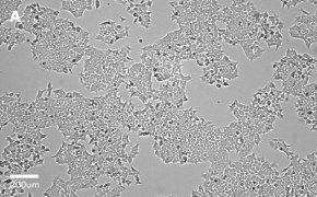
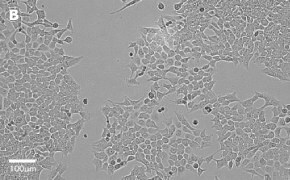
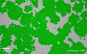
- Images: Brightfield image on Millicell® DCI at 10X (A), 20X (B) and the 10X image with confluency mask (C).
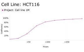
8. Growth Curve
9. Cell culture media:
McCoy’s 5a medium (D6546) + 2 mM L-Glutamine (G7513) + 10% FBS (F2442).
Split sub-confluent cultures (70-80%) 1:3 to 1:10, i.e., seeding at 1-3 x 10,0000 cells/cm2 using 0.25% trypsin or trypsin/EDTA; 5% CO2; 37 °C.
10. Protocols: Cell culture protocols are available in product datasheet.
HEK 293T Cell Line
- Catalog number: 12022001
- Source: Human, fetal kidney cell
- Cell type/morphology: Adherent, small slightly rounded cells growing in a dispersed manner
- Application: Lentiviral particle production, expression of eukaryotic proteins, siRNA transfection, HIV transfection for pseudoviruses production
- Biosafety level: 2
- Handling: Easy
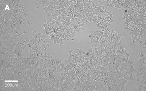
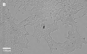
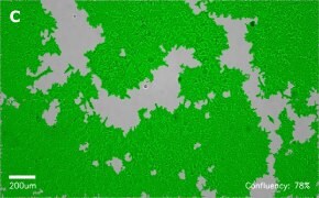
- Images: Brightfield image on Millicell® DCI at 10X (A), 20X (B) and the 10X image with confluency mask (C).
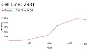
8. Growth Curve
9. Cell culture media:
DMEM-Hi-glucose medium (D6546) + 2 mM L-Glutamine (G7513) + 10% FBS (F2442).
Split sub-confluent cultures (70-80%) 1:4 to 1:10, i.e., seeding at 2-4 x 10,0000 cells/cm2 using 0.25% trypsin or trypsin/EDTA; 5% CO2; 37 °C.
10. Protocols: Cell culture protocols are available in product datasheet.
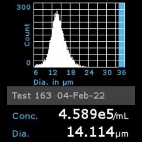
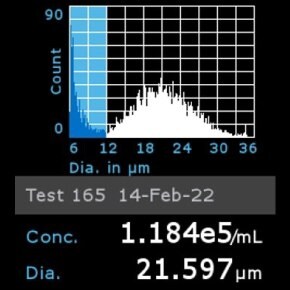
11. Scepter™ histogram: Cells were diluted 1:32 and counted using a Scepter™ 3.0 automated cell counter using 60 µm sensor.
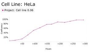
HeLa Cell Line
- Catalog number: 93021013
- Source: Human cervix, epitheloid cervix carcinoma
- Cell type/morphology: Adherent, epithelial
- Application: To study Zika virus (ZIKV) infection; Virus studies; to study the cytotoxicity of extracts, fruits and jams from Sorbus sps
- Biosafety level: 2
- Handling: Easy
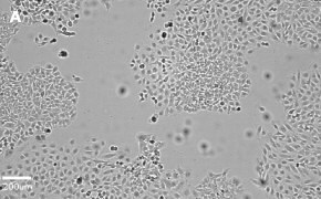
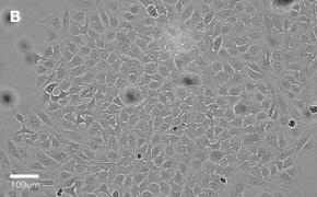
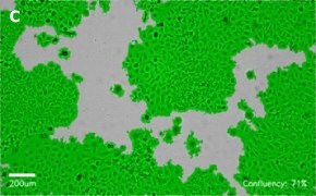
- Images: Brightfield image on Millicell® DCI at 10X (A), 20X (B) and the 10X image with confluency mask (C).
8. Growth Curve
9. Cell culture media:
EMEM (EBSS) + 2 mM L-Glutamine (G7513) + 1% Non-Essential Amino Acids (NEAA) + 10% FBS/FCS (F2442).
Split sub-confluent cultures (70-80%) 1:3 to 1:6, i.e., seeding at 1-3 x 10,0000 cells/cm2 using 0.25% trypsin or trypsin/EDTA; 5% CO2; 37 °C.
10. Protocols: Cell culture protocols are available in product datasheet.
HT29 Cell Line
- Catalog number: 91072201
- Source: Human colon
- Cell type/morphology: Adherent, epithelial
- Application: HT29 Cell Line has been used to determine viral titers of human parechovirus. Study the ability of Bifidobacterium and Lactobacillus strains to counteract the toxic effect of C. difficile LMG21717, Tumorigenicity studies
- Biosafety level: 2
- Handling: Easy
- Images: Brightfield image on Millicell® DCI at 10X (A), 20X (B), and the 10X image with confluency mask (C).


- Growth curve:

- Cell culture media:
McCoy′s5A (M8403) + 2 mM Glutamine (G7513) + 10% Fetal Bovine Serum FBS/FCS (F2442).
Split sub-confluent cultures (70-80%) 1:3 to 1:10, i.e. seeding at 1-3 x 10,000 cells/cm2 using 0.25% Trypsin; 5% CO2; 37°C. - Protocols: Cell culture protocols are available in the product datasheet.
- Scepter™ histogram: Cells were counted on a Scepter™ 3.0 automated cell counter.

Huh-7D 12 Cell Line
- Catalog number: 01042712
- Source: Human liver
- Cell type/morphology: Adherent, epithelial
- Application: Study of intracellular traffic of delta virus antigens and RNA, viral RNA replication and early virus assembly events, and interaction between HDV and HBV
- Biosafety level: 2
- Handling: Easy
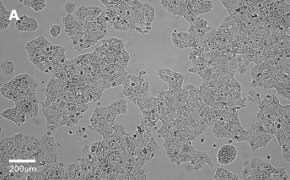
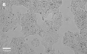
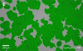
- Images: Brightfield image on Millicell® DCI at 10X (A), 20X (B) and the 10X image with confluency mask (C).
8. Growth Curve
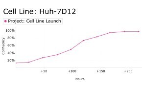
9. Cell culture media:
DMEM-Hi-glucose medium (D6546) + 2 mM L-Glutamine (G7513) + 10% FBS (F2442).
Split sub-confluent cultures (70-80%) 1:3 to 1:6, i.e., seeding at 3-5 x 10,0000 cells/cm2 using 0.25% trypsin or trypsin/EDTA; 5% CO2; 37 °C.
10. Protocols: Cell culture protocols are available in product datasheet.
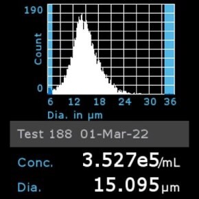
11. Scepter™ histogram: Cells were diluted 1:32 and counted using a Scepter™ 3.0 automated cell counter using 60 µm sensor.
IMR-90 Cell Line
- Catalog number: 85020204
- Source: Human lung (fetal)
- Cell type/morphology: Adherent, fibroblast
- Application: Virus studies, study the cytotoxicity of hydroxyapatite/gold /arginine (HAp/Au/arginine) nanoparticles.
- Biosafety level: 2
- Handling: Easy
- Images: Brightfield image on Millicell® DCI at 10X (A), 20X (B), and the 10X image with confluency mask (C).


- Growth curve:

- Cell culture media:
EMEM (EBSS) (M2279) + 2 mM Glutamine (TMS-002-C) + 1% Non-Essential Amino Acids (NEAA) (TMS-001-C) + 10% Fetal Bovine Serum (FBS) (ES-009-B).
Split sub-confluent cultures (70-80%) 1:3 to 1:6; i.e. seeding at 2-3 x 10,000 cells/cm2 using 0.25% trypsin or trypsin/EDTA; 5% CO2; 37 °C.
- Protocols: Cell culture protocols are available in the product datasheet.
- Scepter™ histogram: Cells were counted on a Scepter™ 3.0 automated cell counter.

J774A.1 Cell Line
- Catalog number: 91051511
- Source: Non-human; mouse blood
- Cell type/morphology: Semi-adherent
- Application: Metabolic studies, test the cytotoxicity of [1,2,3]triazolo[1,5-a]pyridinium salts (having leishmanicidal activity) from triazolopyridines. It has also been used to determine the antitrypanosomatid activity of flavonoid glycosides isolated from Delphinium sps.
- Biosafety level: 2
- Handling: Medium
- Images: Brightfield image on Millicell® DCI at 10X (A), 20X (B), and the 10X image with confluency mask (C).


- Growth curve:

- Cell culture media:
DMEM-Hi-glucose medium (D6429) + 2 mM L-Glutamine (G7513) + 10% FBS (ES-009-B).
Cells are subcultured by harvesting attached and suspension cells. Remove the majority of the medium and centrifuge to harvest cells growing in suspension. Scrape adherent cells remaining in the flask. Resuspend cell pellet from centrifugation and combine with scraped cells. Seed into new flask at 1:3 to 1:4 split ratio, i.e. seeding at 1-3 x 10,000 cells/cm2 Incubate at 37°C; 5% CO2. On receipt, growing order cells may be in suspension. The cells and media should be removed from the flask and spun down to form a pellet. This should then be re-suspended in fresh media and a cell count performed. The cells should then be reseeded at 2-4 x 10⁴/cm and should begin to show signs of re-attachment within 48 hours. - Protocols: Cell culture protocols are available in the product datasheet.
- Scepter™ histogram: Cells were counted on a Scepter™ 3.0 automated cell counter.

L929 Cell Line
- Catalog number: 85011425
- Source: Mouse adipose tissue
- Cell type/morphology: Adherent, fibroblast
- Application: Virus studies: PRV, VSV (Indiana), HSV
- Biosafety level: 2
- Handling: Easy
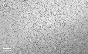
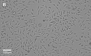
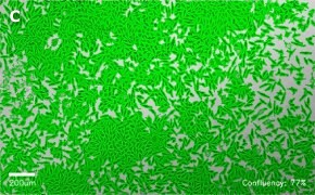
- Images: Brightfield image on Millicell® DCI at 10X (A), 20X (B) and the 10X image with confluency mask (C).
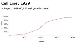
8. Growth Curve
9. Cell culture media:
DMEM-Hi-glucose medium (D6546) + 2 mM L-Glutamine (G7513) + 10% FBS (F2442).
Split sub-confluent cultures (70-80%) 1:3 to 1:6, i.e., seeding at 5 x 1,000-2 X 10,000 cells/cm2 using 0.25% trypsin or trypsin/EDTA; 5% CO2; 37 °C.
10. Protocols: Cell culture protocols are available in product datasheet.
LX-2 Cas9 Cell Line
- Catalog number: SCC613
- Source: Human liver
- Cell type/morphology: Hepatic stellate cells, adherent
- Application: Model of human hepatic fibrosis
- Biosafety level: 2
- Handling: Easy
- Images: Brightfield image on Millicell® DCI at 10X (A) and 10X image with confluency mask (B)


- Genetic profile:

MCF-7 Cell Line
- Catalog number: 86012803
- Source: Human, breast (adenocarcinoma)
- Cell type/morphology: Adherent, epithelial-like
- Application: Tumorigenicity and virus studies: type B and C viruses
- Biosafety level: 2
- Handling: Easy
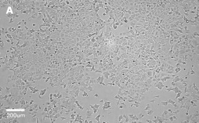
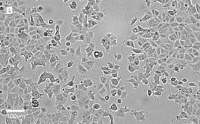
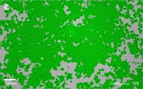
- Images: Brightfield image on Millicell® DCI at 10X (A), 20X (B) and the 10X image with confluency mask (C).
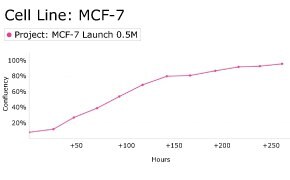
8. Growth Curve
9. Cell culture media:
EMEM (EBSS) + 2 mM L-Glutamine (G7513) + 1% Non-Essential Amino Acids (NEAA) + 10% FBS/FCS (F2442).
Split sub-confluent cultures (70-80%) 1:2 to 1:6, i.e., seeding at 2-4 x 10,0000 cells/cm2 using 0.25% trypsin or trypsin/EDTA; 5% CO2; 37 °C.
10. Protocols: Cell culture protocols are available in product datasheet.
MCF-7 BUS Cell Line
- Catalog number: SCC276
- Source: Human breast cancer
- Cell type/morphology: Adherent, epithelial
- Application: Cellular model for estrogen-responsive metastatic breast cancer
- Biosafety level: 2
- Handling: Easy
- Images: Brightfield image on Millicell® DCI at 10X (A) and 10X image with confluency mask (B)


- Genetic profile:

MDCK Cell Line
- Catalog number: 84121903
- Source: Non-human, Canine kidney
- Cell type/morphology: Adherent, epithelial
- Application: Virus studies: SVEV, VSV, Vaccinia, adeno, and reoviruses
- Biosafety level: 2
- Handling: Easy
- Images: Brightfield image on Millicell® DCI at 10X (A), 20X (B), and the 10X image with confluency mask (C).


- Growth curve:

- Cell culture media:
EMEM (EBSS)(M2279) + 2 mM Glutamine (TMS-002-C) + 1% Non-Essential Amino Acids (NEAA)(TMS-001-C) + 10% Fetal Bovine Serum (FBS)(ES-009-B).
Split sub-confluent cultures (70-80%) 1:3 to 1:10, i.e. seeding at 1-3 x 10,000 cells/cm2 using 0.25% trypsin/EDTA; 5% CO2; 37 °C. - Protocols: Cell culture protocols are available in the product datasheet.
- Scepter™ histogram: Cells were counted on a Scepter™ 3.0 automated cell counter.

MG-63 Cell Line
- Catalog number: 86051601
- Source: Human osteosarcoma
- Cell type/morphology: Adherent, fibroblast
- Application: Interferon-induction studies, to investigate the cytotoxicity of acrylic bone cement extracts on MG-63 cells, to fabricate three-dimensional (3-D) niches and study the cell viability, adhesion, and proliferation of MG-63 cells
- Biosafety level: 2
- Handling: Easy
- Images: Brightfield image on Millicell® DCI at 10X (A), 20X (B), and the 10X image with confluency mask (C).


- Growth curve:

- Cell culture media:
EMEM (EBSS) (M2279) + 2 mM Glutamine (TMS-002-C) + 1% Non-Essential Amino Acids (NEAA) (TMS-001-C) + 10% Fetal Bovine Serum (FBS) (ES-009-B).
Split sub-confluent cultures (70-80%) 1:3 to 1:6, i.e. seeding at 2-4 x 10,000 cells/cm2 using 0.25% trypsin or trypsin/EDTA; 5% CO2; 37 °C. - Protocols: Cell culture protocols are available in the product datasheet.
- Scepter™ histogram: Cells were counted on a Scepter™ 3.0 automated cell counter.

MRC-5 SV2 Cell Line
- Catalog number: 84100401
- Source: Human, fetal lung cell
- Cell type/morphology: Adherent, fibroblast-like fetal lung cell
- Application: Transformation and virus studies: SV40
- Biosafety level: 2
- Handling: Easy
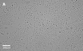
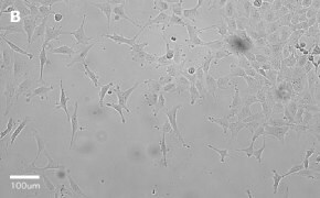
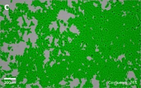
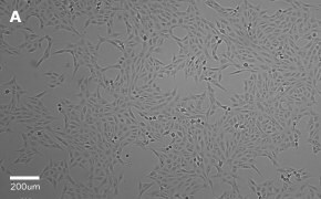
- Images: Brightfield image on Millicell® DCI at 10X (A), 20X (B) and the 10X image with confluency mask (C).
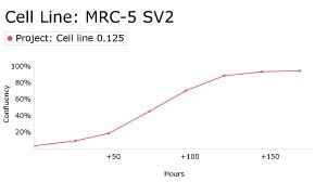
8. Growth Curve
9. Cell culture media:
EMEM (EBSS) + 2 mM L-Glutamine (G7513) + 1% Non-Essential Amino Acids (NEAA) + 10% FBS (F2442).
Split sub-confluent cultures (70-80%) 1:5 to 1:10, i.e., seeding at 1-2 x 10,0000 cells/cm2 using 0.25% trypsin or trypsin/EDTA; 5% CO2; 37 °C.
10. Protocols: Cell culture protocols are available in product datasheet.
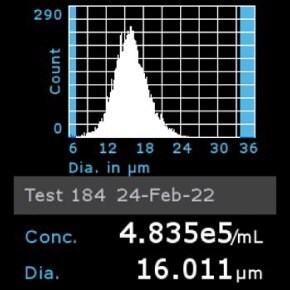
11. Scepter™ histogram: Cells were diluted 1:8 and counted using a Scepter™ 3.0 automated cell counter using 60 µm sensor.
Oli-neu Cell Line
- Catalog number: SCC261
- Source: Non-human, mouse
- Cell type/morphology: Adherent
- Application: neuronal physiology, oligodendrocyte biology
- Biosafety level: 2
- Handling: Advanced
- Images: Brightfield image on Millicell® DCI at 10X (A), 10X image with confluency mask (B), differentiated Oli-neu oligodendrocytes (C)


- Genetic profile:
- Cell culture media:
Cells were thawed and expanded in Oli-neu Expansion Medium in poly-L-lysine-coated tissue culture flasks. Oligo-dendrocyte differentiation was done in expansion medium with daily addition of 1 mM dbcAMP (D0627) - Protocols: Cell culture protocols are available in the product datasheet.
- Scepter™ histogram: Cells were counted on a Scepter™ 3.0 automated cell counter.

OSUMMER.1 Cell Line
- Catalog number: SCC445
- Source: Non-human, male mouse
- Cell type/morphology: Adherent
- Application: Melanoma specific to NRAS mutation Q61R
- Biosafety level: 2
- Handling: Easy
- Images: Brightfield image on Millicell® DCI at 10X (A) and the 10X image with confluency mask (B).


- Genetic profile:
- Cell culture media:
Cells were thawed and expanded in OSUMMER Expansion Medium comprised of DMEM-High Glucose medium (D5796) with 10% FBS (ES-009-B), 2 mM L-Glutamine (TMS-002-C), and 1X penicillin/streptomycin (TMS-AB2-C, optional). - Protocols: Cell culture protocols are available in product datasheet.
- Scepter™ histogram: Cells were counted on a Scepter™ 3.0 automated cell counter.

OSUMMER.2 Cell Line
- Catalog number: SCC446
- Source: Non-human, mouse
- Cell type/morphology: Adherent
- Application: Melanoma specific to NRAS mutation Q61R
- Biosafety level: 2
- Handling: Easy
- Images: Brightfield image on Millicell® DCI at 10X (A) and the 10X image with confluency mask (B).


- Genetic profile:
- Cell culture media:
Cells were thawed and expanded in OSUMMER Expansion Medium comprised of DMEM-High Glucose medium (D5796) with 10% FBS (ES-009-B), 2 mM L-Glutamine (TMS-002-C), and 1X penicillin/streptomycin (TMS-AB2-C, optional). - Protocols: Cell culture protocols are available in product datasheet.
- Scepter™ histogram: Cells were counted on a Scepter™ 3.0 automated cell counter.

OSUMMER.3 Cell Line
- Catalog number: SCC447
- Source: Non-human, female mouse
- Cell type/morphology: Adherent
- Application: Melanoma specific to NRAS mutation Q61R
- Biosafety level: 2
- Handling: Easy
- Images: Brightfield image on Millicell® DCI at 10X (A) and the 10X image with confluency mask (B).


- Genetic profile:
- Cell culture media:
Cells were thawed and expanded in OSUMMER Expansion Medium comprised of DMEM-High Glucose medium (D5796) with 10% FBS (ES-009-B), 2 mM L-Glutamine (TMS-002-C), and 1X penicillin/streptomycin (TMS-AB2-C, optional). - Protocols: Cell culture protocols are available in product datasheet.
- Scepter™ histogram: Cells were counted on a Scepter™ 3.0 automated cell counter.

OSUMMER.4 Cell Line
- Catalog number: SCC448
- Source: Male mouse, melanoma
- Cell type/morphology: Adherent, epithelial
- Application: Carries the NRAS mutation Q61R and may be used to create syngeneic mouse melanoma models
- Biosafety level: 2
- Handling: Easy
- Images: Brightfield image on Millicell® DCI at 10X (A) and the 10X image with confluency mask (B).


- Genetic profile:

OSUMMER.6 Cell Line
- Catalog number: SCC450
- Source: Male mouse, melanoma
- Cell type/morphology: Adherent, epithelial
- Application: Carries the NRAS mutation Q61R and may be used to create syngeneic mouse melanoma models
- Biosafety level: 2
- Handling: Easy
- Images: Brightfield image on Millicell® DCI at 10X (A) and the 10X image with confluency mask (B).


- Genetic profile:

OSUMMER.7 Cell Line
- Catalog number: SCC451
- Source: Male mouse, melanoma
- Cell type/morphology: Adherent, epithelial
- Application: Carries the NRAS mutation Q61R and may be used to create syngeneic mouse melanoma models
- Biosafety level: 2
- Handling: Easy
- Images: Brightfield image on Millicell® DCI at 10X (A) and the 10X image with confluency mask (B).


- Genetic profile:

OSUMMER.8 Cell Line
- Catalog number: SCC452
- Source: Non-human, female mouse
- Cell type/morphology: Adherent
- Application: Melanoma specific to NRAS mutation Q61K
- Biosafety level: 2
- Handling: Easy
- Images: Brightfield image on Millicell® DCI at 10X (A) and the 10X image with confluency mask (B).


- Genetic profile:
- Cell culture media:
Cells were thawed and expanded in OSUMMER Expansion Medium comprised of DMEM-High Glucose medium (D5796) with 10% FBS (ES-009-B), 2 mM L-Glutamine (TMS-002-C), and 1X penicillin/streptomycin (TMS-AB2-C, optional). - Protocols: Cell culture protocols are available in product datasheet.
- Scepter™ histogram: Cells were counted on a Scepter™ 3.0 automated cell counter.

OSUMMER.9 Cell Line
- Catalog number: SCC453
- Source: Male mouse, melanoma
- Cell type/morphology: Adherent, epithelial
- Application: Carries the NRAS mutation Q61L and may be used to create syngeneic mouse melanoma models
- Biosafety level: 2
- Handling: Easy
- Images: Brightfield image on Millicell® DCI at 10X (A) and the 10X image with confluency mask (B).


- Genetic profile:

OSUMMER.10 Cell Line
- Catalog number: SCC454
- Source: Non-human, female mouse
- Cell type/morphology: Adherent
- Application: Melanoma specific to NRAS mutation Q13R
- Biosafety level: 2
- Handling: Easy
- Images: Brightfield image on Millicell® DCI at 10X (A) and the 10X image with confluency mask (B).


- Genetic profile:
- Cell culture media:
Cells were thawed and expanded in OSUMMER Expansion Medium comprised of DMEM-High Glucose medium (D5796) with 10% FBS (ES-009-B), 2 mM L-Glutamine (TMS-002-C), and 1X penicillin/streptomycin (TMS-AB2-C, optional). - Protocols: Cell culture protocols are available in product datasheet.
- Scepter™ histogram: Cells were counted on a Scepter™ 3.0 automated cell counter.

OSUMMER.11 Cell Line
- Catalog number: SCC455
- Source: Non-human, female mouse
- Cell type/morphology: Adherent
- Application: Melanoma specific to NRAS mutation Q61R
- Biosafety level: 2
- Handling: Easy
- Images: Brightfield image on Millicell® DCI at 10X (A) and the 10X image with confluency mask (B).


- Genetic profile:
- Cell culture media:
Cells were thawed and expanded in OSUMMER Expansion Medium comprised of DMEM-High Glucose medium (D5796) with 10% FBS (ES-009-B), 2 mM L-Glutamine (TMS-002-C), and 1X penicillin/streptomycin (TMS-AB2-C, optional). - Protocols: Cell culture protocols are available in product datasheet.
- Scepter™ histogram: Cells were counted on a Scepter™ 3.0 automated cell counter.

Scp2 Cell Line
- Catalog number: SCC458
- Source: Non-human, mouse
- Cell type/morphology: Adherent, epithelial
- Application: Mammary epithelium biology
- Biosafety level: 2
- Handling: Easy
- Images: Brightfield image on Millicell® DCI at 10X (A) and the 10X image with confluency mask (B).


- Genetic profile:
- Cell culture media:
Cells were thawed and expanded in Scp2 Expansion Medium comprised of DMEM/F12-High medium (D8062) with 2% FBS (ES-009-B), 2 mM L-Glutamine (TMS-002-C), 5 mg/mL insulin (I9278), and 1X penicillin/streptomycin (TMS-AB2-C, optional). - Protocols: Cell culture protocols are available in product datasheet.
- Scepter™ histogram: Cells were counted on a Scepter™ 3.0 automated cell counter.

T24T Cell Line
- Catalog number: SCC468
- Source: Human bladder carcinoma
- Cell type/morphology: Adherent, epithelial
- Application: To study metastatic bladder cancer
- Biosafety level: 2
- Handling: Easy
- Images: Brightfield image on Millicell® DCI at 10X (A) and 10X image with confluency mask (B)


- Genetic profile:

Vero Cell Line
- Catalog number: 84113001
- Source: Non-human, African Green Monkey
- Cell type/morphology: Adherent, fibroblast-like kidney cell
- Application: Media testing, Toxicology, Bioproduction, 3D cell culture
- Biosafety level: 2
- Handling: Easy
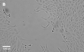
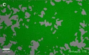
- Images: Brightfield image on Millicell® DCI at 10X (A), 20X (B) and the 10X image with confluency mask (C).
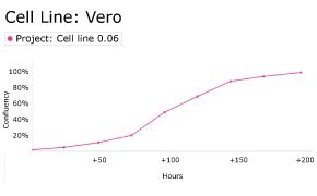
8. Growth Curve
9. Cell culture media:
DMEM-Hi-glucose medium (D6546) + 2 mM L-Glutamine (G7513) + 10% FBS (F2442).
Split sub-confluent cultures (70-80%) 1:3 to 1:6 using 0.25% trypsin or trypsin/EDTA; 5% CO2; 37 °C.
10. Protocols: Cell culture protocols are available in product datasheet.
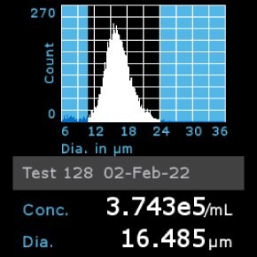
11. Scepter™ histogram: Cells were counted using a Scepter™ 3.0 automated cell counter.
Vero C1008 Cell Line
- Catalog number: 85020206
- Source: Non-human, African Green Monkey [Vero 76, clone E6, Vero E6]
- Cell type/morphology: Adherent, fibroblast-like kidney cell
- Application: Media testing, Toxicology, Bioproduction, 3D cell culture
- Biosafety level: 2
- Handling: Easy
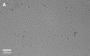
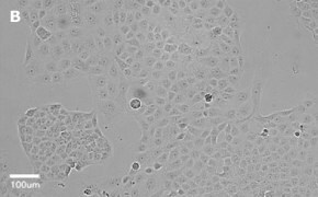
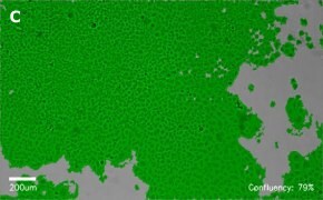
- Images: Brightfield image on Millicell® DCI at 10X (A), 20X (B) and the 10X image with confluency mask (C).
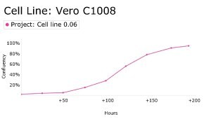
8. Growth Curve
9. Cell culture media:
DMEM-Hi-glucose medium (D6546) + 2 mM L-Glutamine (G7513) + 10% FBS (F2442).
Split sub-confluent cultures (70-80%) 1:3 to 1:6 using 0.25% trypsin or trypsin/EDTA; 5% CO2; 37 °C.
10. Protocols: Cell culture protocols are available in product datasheet.
11. Scepter™ histogram: Cells were diluted 1:16 and counted using a Scepter™ 3.0 automated cell counter using 60 µm sensor.
WI 38 Cell Line
- Catalog number: 90020107
- Source: Human, Female Caucasian, fetal lung cell
- Cell type/morphology: Adherent, fibroblast-like fetal lung cell
- Application: Virus studies (rhinoviruses), vaccine production, study cytotoxicity of acrylic based resin compounds and silica nanoparticles
- Biosafety level: 2
- Handling: Easy
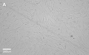
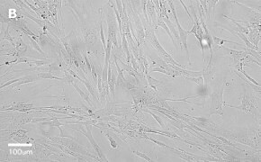
- Images: Brightfield image on Millicell® DCI at 10X (A), 20X (B) and the 10X image with confluency mask (C).

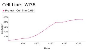
8. Growth Curve
9. Cell culture media:
EMEM (EBSS) + 2 mM L-Glutamine (G7513) + 1% Non-Essential Amino Acids (NEAA) + 15% FBS (F2442).
Split sub-confluent cultures (70-80%) 1:3 to 1:6, i.e., seeding at 2-4 x 10,0000 cells/cm2 using 0.25% trypsin or trypsin/EDTA; 5% CO2; 37 °C.
Finite lifespan, 50 ± 10 population doublings.
10. Protocols: Cell culture protocols are available in product datasheet.
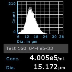
11. Scepter™ histogram: Cells were diluted 1:8 and counted using a Scepter™ 3.0 automated cell counter using 60 µm sensor.
To continue reading please sign in or create an account.
Don't Have An Account?