
05-915
Anti-N-Cadherin Antibody, clone 13A9
culture supernatant, clone 13A9, Upstate®
Sinônimo(s):
Cadherin-2, CD325, CDw325, N-cadherin, Neural cadherin
Selecione um tamanho
Selecione um tamanho
About This Item
Produtos recomendados

fonte biológica
mouse
Nível de qualidade
forma do anticorpo
culture supernatant
tipo de produto de anticorpo
primary antibodies
clone
13A9, monoclonal
reatividade de espécies
human
embalagem
antibody small pack of 25 μL
fabricante/nome comercial
Upstate®
técnica(s)
immunocytochemistry: suitable
immunohistochemistry: suitable
immunoprecipitation (IP): suitable
western blot: suitable
nº de adesão NCBI
Descrição geral
Especificidade
Imunogênio
Aplicação
Immunohistochemistry Analysis: A representative lot detected strong N-cadherin immunoreactivity in paraffin-embedded rectal cancer (RC) tissues with positive regional lymph node metastasis (RLNM) status, while only weak N-cadherin immunoreactivity was detected in RC with negative RLNM, and no N-cadherin staining was seen in normal colorectal epithelium (Fan, X.J., et al. (2012). Br. J. Cancer. 106(11):1735-1741).
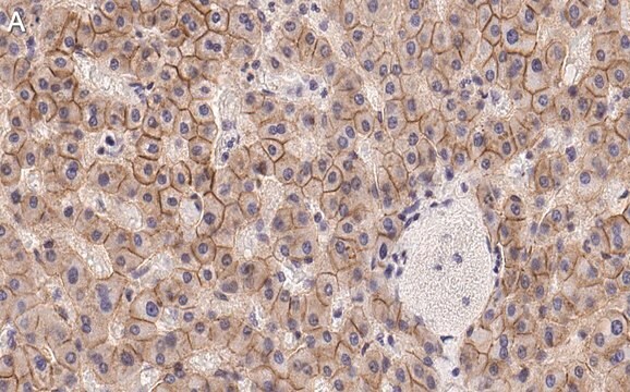
Immunohistochemistry Analysis: A representative lot detected N-cadherin immunoreactivity in formalin-fixed, paraffin-embedded hepatocellular carcinoma (HCC) tissue sections. A significant inverse correlation was found between RUNX3 and N-cadherin expression levels (Tanaka, S., et al. (2012). Int. J. Cancer. 131(11):2537-2546).
Western Blotting Analysis: A representative lot detected an upregulated N-cadherin expression in CCL185 carcinoma cells following transient Epstein-Barr virus (EBV) infection. The EMT-like phenotype remained even after viral loss by culture selection pressure withdrawal (Queen, K.J., et al. (2013). Int. J. Cancer. 132(9):2076-2086).
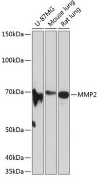
Western Blotting Analysis: A representative lot detected N-cadherin in Hep3B, Huh7, HLF and SK-Hep1 human hepatocellular carcinoma (HCC) cell lysates (Tanaka, S., et al. (2012). Int. J. Cancer. 131(11):2537-2546).
Western Blotting Analysis: A representative lot detected both the unprocessed (pro-) and processed (mature) forms of N-cadherin in HeLa cell lysate (Wahl, J.K. 3rd., et al. (2003). J. Biol. Chem. 278(19):17269-17276).
Western Blotting Analysis: A representative lot detected N-cadherin in WI-38 human fibroblast lysate, but not in JAr human placental choriocarcinoma cell lysate (Knudsen, K.A., et al. (1995). J. Cell Biol. 130(1):67-77).
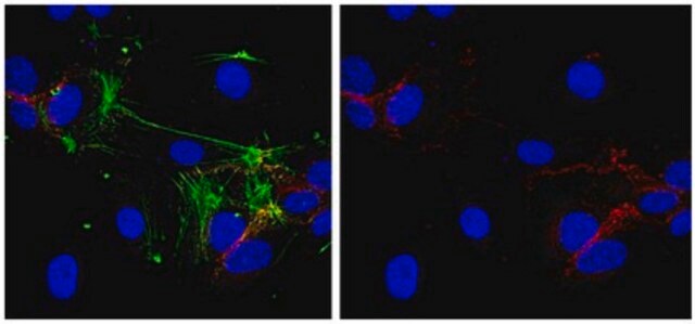
Immunocytochemistry Analysis: A representative lot detected N-cadherin immunoreactivity localized primarily at the cell-cell borders by fluorescent immunocytochemistry staining of 1% paraformaldehyde-fixed, methanol-permeabilized HeLa cells (Wahl, J.K. 3rd., et al. (2003). J. Biol. Chem. 278(19):17269-17276).
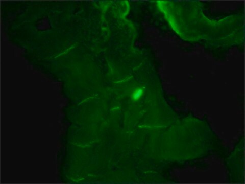
Immunocytochemistry Analysis: A representative lot detected N-cadherin immunoreactivity colocalized with those of alpha- and beta-catenin by dual fluorescent immunocytochemistry staining of fixed WI-38 human fibroblasts (Knudsen, K.A., et al. (1995). J. Cell Biol. 130(1):67-77).
Immunoprecipitation Analysis: Representative lots co-immunoprecipitated alpha-catenin, beta-catenin, and plakoglobin with N-cadherin from WI-38 human fibroblast and HeLa cell lysates (Wahl, J.K. 3rd., et al. (2003). J. Biol. Chem. 278(19):17269-17276; Knudsen, K.A., et al. (1995). J. Cell Biol. 130(1):67-77).
Cell Structure
Adhesion (CAMs)
Qualidade
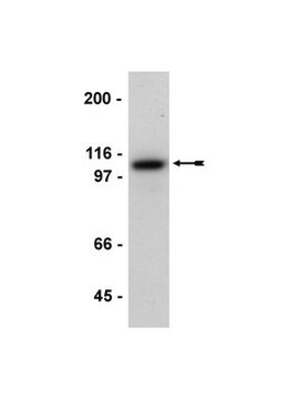
Western Blotting Analysis: A 1:1000-5000 dilution of this hybridoma culture supernatant detected N-cadherin in HeLa cell lysate.
Descrição-alvo
Ligação
forma física
Armazenamento e estabilidade
Nota de análise
HeLa cell lysate
Informações legais
Exoneração de responsabilidade
Não está encontrando o produto certo?
Experimente o nosso Ferramenta de seleção de produtos.
recomendado
Certificados de análise (COA)
Busque Certificados de análise (COA) digitando o Número do Lote do produto. Os números de lote e remessa podem ser encontrados no rótulo de um produto após a palavra “Lot” ou “Batch”.
Já possui este produto?
Encontre a documentação dos produtos que você adquiriu recentemente na biblioteca de documentos.
Artigos
Human iPSC neural differentiation media and protocols used to generate neural stem cells, neurons and glial cell types.
Nossa equipe de cientistas tem experiência em todas as áreas de pesquisa, incluindo Life Sciences, ciência de materiais, síntese química, cromatografia, química analítica e muitas outras.
Entre em contato com a assistência técnica