Skip to Content
Merck
HomeProtein Labeling & ModificationPeptide Modifications: N-Terminal, Internal, and C-Terminal

Peptide Modifications: N-Terminal, Internal, and C-Terminal

N-terminal, internal, and C-terminal peptide modifications are useful for a variety of applications, such as Western blotting, protein-protein interaction studies, and fluorescence-based assays. Use the table below for a list of various applications and to jump to more information, including structures and relevant references.

To request a quote on custom peptides or for help designing a custom PEPscreen® peptide library, follow the link below.

Peptide ModificationApplications
AcetylationIncrease peptide stability by preventing N-terminal degradation
BiotinCommonly used in immunoassays, histocytochemistry, and fluorescence based flow cytometry
Dansyl and 2, 4-DinitrophenylFluorescence based assays
Fluorescein and 7-methoxycoumarin acetic acidProtein-protein interaction and localization studies
Palmitic AcidIncrease their cell permeability and help binding of the peptides to cell membrane
Cyclization (Disulfide bridge)Stabilize the peptide conformation, increase bioactivity, and enzyme stability
Cysteine Carbamidomethylation (CAM)Peptide Mass Fingerprinting for identification and characterization of peptides. To block cysteine residues from oxidation in protein assays
PhosphorylationGene expression, protein-protein interaction and signal transduction in plants and animals
Isotope labeled Amino AcidsStudy protein interactions, proteins, post translation modifications such as ubiquitination and phosphorylation
SpacersReduce steric hindrance at the binding sites of the peptide and its load

1.0 N-Terminal Modifications

1.1 Acetylation

This modification removes the positive charge on the N-terminal of peptides, thus mimicking natural proteins. In some cases, it increases peptide stability by preventing N-terminal degradation 1-2

Acetylation
Molecular Weight43 g/mol
Availability:PEPscreen®

References

1.
Arnesen T. Towards a Functional Understanding of Protein N-Terminal Acetylation. PLoS Biol. 9(5):e1001074. https://doi.org/10.1371/journal.pbio.1001074
2.
Wallace RJ. 1992. Acetylation of peptides inhibits their degradation by rumen micro-organisms. Br J Nutr. 68(2):365-372. https://doi.org/10.1079/bjn19920095

1.2 Biotin

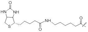
Biotin has a very strong affinity for streptavidin and avidin. Biotin-labeled peptides are commonly used in immunoassays1, histocytochemistry2, and fluorescence based flow cytometry3

Biotin
Molecular Weight340 g/mol
Availability:PEPscreen®

References

1.
Sélo I, Négroni L, Créminon C, Grassi J, Wal J. 1996. Preferential labeling of ?-amino N-terminal groups in peptides by biotin: application to the detection of specific anti-peptide antibodies by enzyme immunoassays. Journal of Immunological Methods. 199(2):127-138. https://doi.org/10.1016/s0022-1759(96)00173-1
2.
HOWL J, WANG X, KIRK CJ, WHEATLEY M. 1993. Fluorescent and biotinylated linear peptides as selective bifunctional ligands for the V1a vasopressin receptor. Eur J Biochem. 213(2):711-719. https://doi.org/10.1111/j.1432-1033.1993.tb17811.x
3.
Buranda, et. al. 1991. Peptide, antibodies and FRET on beads in flow cytometry: a model system using fluoresceinated and biotinylated β-endorphin. Cytometry . 3721-31.

1.3 Dansyl

Dansyl labeled peptides are used in fluorescence based assays.

Dansyl
Molecular Weight249 g/mol
Excitation Wavelength372 nm
Availability:PEPscreen®

References

1.
Glukhov E, Stark M, Burrows LL, Deber CM. 2005. Basis for Selectivity of Cationic Antimicrobial Peptides for BacterialVersusMammalian Membranes. J. Biol. Chem.. 280(40):33960-33967. https://doi.org/10.1074/jbc.m507042200
2.
Matsuzaki K, Mitani Y, Akada K, Murase O, Yoneyama S, Zasloff M, Miyajima K. 1998. Mechanism of Synergism between Antimicrobial Peptides Magainin 2 and PGLa?. Biochemistry. 37(43):15144-15153. https://doi.org/10.1021/bi9811617
3.
Pecht I, Maron E, Arnon R, Sela M. 1971. Specific Excitation Energy Transfer from Antibodies to Dansyl-Labeled Antigen. Studies with the "Loop" Peptide of Hen Egg-White Lysozyme. Eur J Biochem. 19(3):368-371. https://doi.org/10.1111/j.1432-1033.1971.tb01325.x

1.4 2, 4-Dinitrophenyl

2, 4-DNP is used as a quencher for (7-methoxy coumarin-4-yl) acetyl (MCA) and sometimes tryptophan. This modification can be attached at the N-terminal of a peptide or as an internal modification through lysine side chain.

Dinitrophenyl
olecular Weight167 g/mol
Excitation Wavelength354-400 nm
Availability:PEPscreen®

References

1.
Vickers C, Hales P, Kaushik V, Dick L, Gavin J, Tang J, Godbout K, Parsons T, Baronas E, Hsieh F, et al. 2002. Hydrolysis of Biological Peptides by Human Angiotensin-converting Enzyme-related Carboxypeptidase. J. Biol. Chem.. 277(17):14838-14843. https://doi.org/10.1074/jbc.m200581200
2.
Knight C, Willenbrock F, Murphy G. 1992. A novel coumarin-labelled peptide for sensitive continuous assays of the matrix metalloproteinases. 296(3):263-266. https://doi.org/10.1016/0014-5793(92)80300-6

1.5 Fluorescein

Fluorescein-labeled peptides have many fluorescein based biomolecular applications including protein-protein interaction, flow cytometry and localization studies1-3.

Fluorescein
Molecular Weight359 g/mol
Excitation/Emission Wavelength494/518 nm
Availability:PEPscreen®

References

1.
Richard JP, Melikov K, Vives E, Ramos C, Verbeure B, Gait MJ, Chernomordik LV, Lebleu B. 2003. Cell-penetrating Peptides. J. Biol. Chem.. 278(1):585-590. https://doi.org/10.1074/jbc.m209548200
2.
Farley RA, Tran CM, Carilli CT, Hawke D, Shively JE. 1984. The amino acid sequence of a fluorescein-labeled peptide from the active site of (Na,K)-ATPase. J Biol Chem. 259(15):9532-5.
3.
Futaki S, Suzuki T, Ohashi W, Yagami T, Tanaka S, Ueda K, Sugiura Y. 2001. Arginine-rich Peptides. J. Biol. Chem.. 276(8):5836-5840. https://doi.org/10.1074/jbc.m007540200
4.
Foerg C, Weller KM, Rechsteiner H, Nielsen HM, Fernández-Carneado J, Brunisholz R, Giralt E, Merkle HP. 2008. Metabolic Cleavage and Translocation Efficiency of Selected Cell Penetrating Peptides: A Comparative Study with Epithelial Cell Cultures. AAPS J. 10(2):349-359. https://doi.org/10.1208/s12248-008-9029-4

1.6 7-methoxycoumarin acetic acid (Mca)

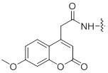
7-methoxy coumarin-labeled peptides have applications in protein-protein interaction and localization studies1-3.

7-methoxycoumarin acetic acid (Mca)
Molecular Weight217 g/mol
Excitation/Emission Wavelength323/382 nm
Availability:PEPscreen®

References

1.
Vidal, et. al. 1996. Solid-Phase Synthesis and Cellular Localization of a C-and/or N-terminal Labeled Peptide. Journal of Peptide Science. 2125-133.
2.
Yandek LE, Pokorny A, Florén A, Knoelke K, Langel Ü, Almeida PF. 2007. Mechanism of the Cell-Penetrating Peptide Transportan 10 Permeation of Lipid Bilayers. Biophysical Journal. 92(7):2434-2444. https://doi.org/10.1529/biophysj.106.100198

1.7 Palmitic Acid

Palmitic acid is 16-carbon fatty acid that is conjugated to peptides to increase their cell permeability and help binding of the peptides to cell membrane 1-2.

Palmitic Acid
Molecular Weight239 g/mol
Availability:PEPscreen®

References

1.
Avrahami D, Shai Y. 2004. A New Group of Antifungal and Antibacterial Lipopeptides Derived from Non-membrane Active Peptides Conjugated to Palmitic Acid. J. Biol. Chem.. 279(13):12277-12285. https://doi.org/10.1074/jbc.m312260200
2.
Buss JE, Sefton BM. 1986. Direct identification of palmitic acid as the lipid attached to p21ras.. Mol. Cell. Biol.. 6(1):116-122. https://doi.org/10.1128/mcb.6.1.116
Molecular WeightN/A
Availability:PEPscreen®, AQUA™ Peptides

References

1.
Góngora-Benítez M, Tulla-Puche J, Albericio F. 2014. Multifaceted Roles of Disulfide Bonds. Peptides as Therapeutics. Chem. Rev.. 114(2):901-926. https://doi.org/10.1021/cr400031z
2.
Schmelz EA, Huffaker A, Carroll MJ, Alborn HT, Ali JG, Teal PE. 2012. An Amino Acid Substitution Inhibits Specialist Herbivore Production of an Antagonist Effector and Recovers Insect-Induced Plant Defenses. Plant Physiol.. 160(3):1468-1478. https://doi.org/10.1104/pp.112.201061

2.2 Cysteine Carbamidomethylation (CAM)

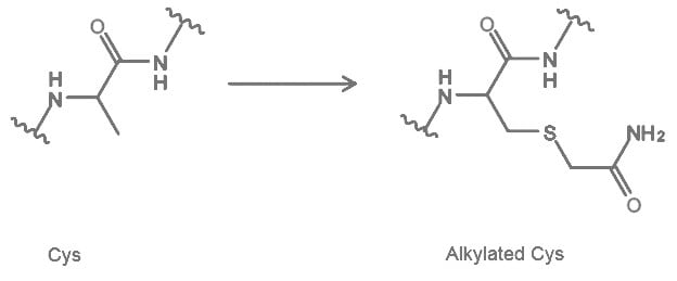
Carbamidomethylation (CAM) is a deliberate post-translational modification introduced to cysteine residues by reacting with iodoacetamide. Peptides with this modification are mainly used in Peptide Mass Fingerprinting for identification and characterization of proteins1. In other assays, this process is used to block Cysteine from oxidation2.
Cysteine Carbamidomethylation (CAM)
Molecular Weight160 g/mol
Availability:PEPscreen®, AQUA™ Peptides

References

1.
Wilkins MR, Appel RD, Williams KL, Hochstrasser DF. 2007. Proteome Research. https://doi.org/10.1007/978-3-540-72910-5
2.
Rombouts I, Lagrain B, Brunnbauer M, Delcour JA, Koehler P. 2013. Improved identification of wheat gluten proteins through alkylation of cysteine residues and peptide-based mass spectrometry. Sci Rep. 3(1): https://doi.org/10.1038/srep02279

2.3 Isotope labeled Amino Acids

AQUA peptides are synthetic peptides with amino acids enriched in 18O, 13C, and/or 14N. They are similar to their native peptides in terms of chemical, physical properties and also their biological activities1. Main applications for these peptides are to study protein interactions, proteins, post translation modifications such as ubiquitination and phosphorylation2-5.

References

1.
Kettenbach AN, Rush J, Gerber SA. 2011. Absolute quantification of protein and post-translational modification abundance with stable isotope?labeled synthetic peptides. Nat Protoc. 6(2):175-186. https://doi.org/10.1038/nprot.2010.196
2.
Li H, Xu C, Blais S, Wan Q, Zhang H, Landry SJ, Hioe CE. 2009. Proximal Glycans Outside of the Epitopes Regulate the Presentation of HIV-1 Envelope gp120 Helper Epitopes. J Immunol. 182(10):6369-6378. https://doi.org/10.4049/jimmunol.0804287
3.
Le Bihan T, Grima R, Martin S, Forster T, Le Bihan Y. 2010. Quantitative analysis of low-abundance peptides in HeLa cell cytoplasm by targeted liquid chromatography/mass spectrometry and stable isotope dilution: emphasising the distinction between peptide detection and peptide identification. Rapid Commun. Mass Spectrom.. 24(7):1093-1104. https://doi.org/10.1002/rcm.4487
4.
Santhoshkumar P, Raju M, Sharma KK. ?A-Crystallin Peptide 66SDRDKFVIFLDVKHF80 Accumulating in Aging Lens Impairs the Function of ?-Crystallin and Induces Lens Protein Aggregation. PLoS ONE. 6(4):e19291. https://doi.org/10.1371/journal.pone.0019291
5.
Sato Y, Miyashita A, Iwatsubo T, Usui T. 2012. Simultaneous Absolute Protein Quantification of Carboxylesterases 1 and 2 in Human Liver Tissue Fractions using Liquid Chromatography-Tandem Mass Spectrometry. Drug Metab Dispos. 40(7):1389-1396. https://doi.org/10.1124/dmd.112.045054
6.
Brun V, Masselon C, Garin J, Dupuis A. 2009. Isotope dilution strategies for absolute quantitative proteomics. Journal of Proteomics. 72(5):740-749. https://doi.org/10.1016/j.jprot.2009.03.007

2.4 Phosphorylation

Phosphorylation can be performed on Tyr, Ser and Thr residues as a posttranslational modification (PTM) on peptides. Phosphorylated peptides have application in many cellular processes such as gene expression, protein-protein interaction and signal transduction in plants and animals1,2.

Phosphorylation
Molecular Weight82 g/mol
Availability:PEPscreen®, AQUA™ Peptides

References

1.
Guenther JF, Chanmanivone N, Galetovic MP, Wallace IS, Cobb JA, Roberts DM. 2003. Phosphorylation of Soybean Nodulin 26 on Serine 262 Enhances Water Permeability and Is Regulated Developmentally and by Osmotic Signals. Plant Cell. 15(4):981-991. https://doi.org/10.1105/tpc.009787

2.5 Spacers

Spacers are used to create a distance between the peptide and the cargo to reduce steric hindrance at the binding sites of the peptide. In this case cargo can be a drug, dye, tag.

2.5.1 PEGylation

Attachment of poly (ethylene glycol) to a peptide is called PEGylation. Short bifunctional PEG (Poly (ethylene glycol)) can be used as a spacer in bioconjugation of peptides with other molecules. PEG bioconjugation has also been used to improve proteolytic stability, biodistribution and solubility of peptides1-2.

PEGylation
Molecular Weight146 g/mol
Availability:PEPscreen®

References

1.
Sullivan TP, van Poll ML, Dankers PYW, Huck WTS. 2004. Forced Peptide Synthesis in Nanoscale Confinement under Elastomeric Stamps. Angew. Chem.. 116(32):4286-4289. https://doi.org/10.1002/ange.200460271
2.
Veronese FM. 2001. Peptide and protein PEGylation. Biomaterials. 22(5):405-417. https://doi.org/10.1016/s0142-9612(00)00193-9

2.5.2 Amino hexanoic acid

Amino hexanoic acid is a hydrophobic spacer to which a molecule- either a fluorophore, tag or any biological molecule - can be attached to a peptide 1-2.

Amino hexanoic acid
Molecular Weight113 g/mol
Availability:PEPscreen®

Reference

1.
Mitchell D, Steinman L, Kim D, Fathman C, Rothbard J. 2000. Polyarginine enters cells more efficiently than other polycationic homopolymers. J Pept Res. 56(5):318-325. https://doi.org/10.1034/j.1399-3011.2000.00723.x

3.0 C-Terminal Modifications

3.1 Amide (Amidation)

The C-terminal of the peptide is synthesized as an amide to neutralize negative charge created by the C-terminal COOH. This modification is added to prevent enzyme degradation, to mimic native proteins, and in some cases to remove hydrogen bonding at the C-terminal of the peptides which may interfere with the assays.1

Reference

1.
Kim K, Seong BL. 2001. Peptide amidation: Production of peptide hormonesin vivo andin vitro. Biotechnol. Bioprocess Eng.. 6(4):244-251. https://doi.org/10.1007/bf02931985
Sign In To Continue

To continue reading please sign in or create an account.

Don't Have An Account?