General description
Monoclonal Anti-Human Fas (CD95/Apo-1) (mouse IgG1 isotype) is derived from the DX2 hybridoma produced by the fusion of mouse myeloma cells and splenocytes from C3H mice immunized with murine L cells transfected by a human Fas/CD95 cDNA. CD95/Fas/Apo-1 exhibits strong homologies with the extracellular domain of receptors belonging to the tumor necrosis factor (TNF) receptor family, which includes TNF receptor types 1 and 2 (TNFR1/2), the low affinity nerve growth factor receptor, and lymphocyte receptors such as CD27, CD30, CD40, and OX40. Fas is an integral membrane protein, with strong homology to TNF-α and -β , has been identified as Fas ligand.
Many cells can be activated to undergo apoptosis following the interaction of selected ligands with cell surface receptors. The most well studied receptors are CD95/Fas/Apo-1 (apoptosis inducing protein 1) and tumor necrosis factor receptor 1 (TNFR1). Apoptosis mediated by either of these results in activation of the caspases. However, Fas-mediated death occurs much more rapidly than that triggered by TNFR1. Human Fas/CD95/Apo-1 is a single transmembrane glycoprotein receptor (325 amino acids, 45-48 kDa).
Specificity
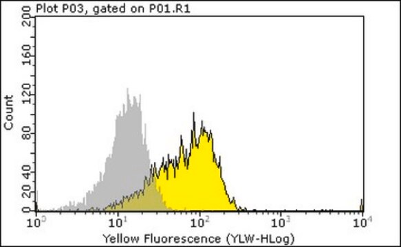
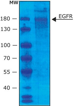
Reacts specifically with the functional epitope of human Fas (CD95/Apo-1) antigen. By immunoblotting, the clone recognizes denatured, non-reduced recombinant human Fas (amino acid residues 1-173). The antibody is reactive in flow cytometry, and may be reactive in the induction of apoptosis.
Immunogen
murine L cells transfected with a human Fas/CD95 cDNA.
Application
Applications in which this antibody has been used successfully, and the associated peer-reviewed papers, are given below.
Western Blotting (1 paper)Monoclonal Anti-Fas (CD95/Apo-1) antibody is suitable for apoptosis treatment of Jurkat cells to study the contribution of channel type into the net K+ flux. It is also suitable for flow cytometry at a concentration of 4-20μg/mL using cultured human Burkitt′s lymphoma Raji cells.
Monoclonal Anti-Fas (CD95/Apo-1) antibody produced in mouse has been used in:
- immunoblotting
- flow cytometry
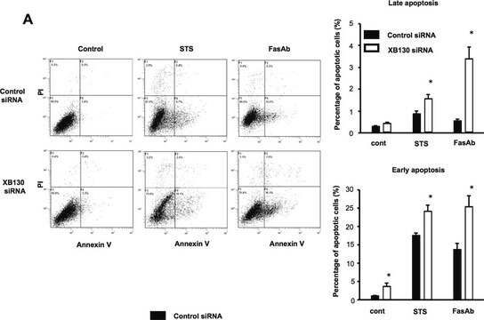
- the induction of apoptosis
Biochem/physiol Actions
Fas is expressed in a number of lymphoma cell lines, on Epstein-Barr virus-transformed B lymphoblasts and on a proportion of activated B and T cells. Fas is also detected in soluble form and this form of the protein is thought to play a role in regulating certain aspects of immune system function. Elevated levels of soluble Fas is observed in leukemia and systemic lupus erythematosus. Therefore, altered levels of secreted Fas protein is likely to be involved in the abnormal growth regulation of lymphoid cells.
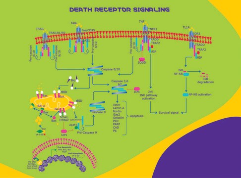
Human CD95/Fas/Apo-1 antigen is a single transmembrane glycoprotein receptor of 325 amino acids (45-48 kDa) which activate cell apoptosis. The action of Fas is mediated via FADD (Fas-associated death domain)/ MORT1, an adapter protein that has a death domain at its C-terminus and binds to the cytoplasmic death domain of Fas. APO-1/Fas(CD95) comprises of a death domain (DD) within the cytoplasmic region which triggers apoptosis upon binding of their cognate ligands. Once it is activated, APO-1/Fas(CD95) further aggregates its intracellular death domains which leads to the recruitment of two key signaling proteins followed by the formation of death-inducing signaling complex. These complex crosslinks through its C-terminal DD with APO-1/Fas receptors and engage caspase-8 via its N-terminal death effector domain (DED) to the DISC.
Physical form
Solution from a bioreactor culture supernatant in 0.01 M phosphate buffered saline, pH 7.4, containing 15 mM sodium azide.
Disclaimer
Unless otherwise stated in our catalog or other company documentation accompanying the product(s), our products are intended for research use only and are not to be used for any other purpose, which includes but is not limited to, unauthorized commercial uses, in vitro diagnostic uses, ex vivo or in vivo therapeutic uses or any type of consumption or application to humans or animals.
