Direkt zum Inhalt
Merck
Alle Fotos(4)

Dokumente

MABT1340

Sigma-Aldrich

Anti-Lamin A/C Antibody, clone 2A1

clone 2A1, from mouse

Synonym(e):

70 kDa Lamin, Renal carcinoma antigen NY-REN-32

Anmeldenzur Ansicht organisationsspezifischer und vertraglich vereinbarter Preise


About This Item

UNSPSC-Code:
12352203
eCl@ss:
32160702
NACRES:
NA.41

Biologische Quelle

mouse

Antikörperform

purified immunoglobulin

Antikörper-Produkttyp

primary antibodies

Klon

2A1, monoclonal

Speziesreaktivität

mouse, monkey, hamster, human

Verpackung

antibody small pack of 25 μg

Methode(n)

immunocytochemistry: suitable
immunofluorescence: suitable
immunoprecipitation (IP): suitable
western blot: suitable

Isotyp

IgG2bκ

NCBI-Hinterlegungsnummer

UniProt-Hinterlegungsnummer

Posttranslationale Modifikation Target

unmodified

Angaben zum Gen

human ... LMNA(4000)

Allgemeine Beschreibung

Prelamin-A/C (UniProt: P02545) is encoded by the LMNA (also known as LMN1) gene (Gene ID: 4000) in human. It is cleaved into Lamin-A/C (also known as 70 kDa Lamin, Renal carcinoma antigen NY-REN-32). Lamins are components of the nuclear lamina that provide a framework for the nuclear envelope and may also interact with chromatin. Lamin A and C are present in equal amounts in the lamina of mammals. Plays an important role in nuclear assembly, chromatin organization, nuclear membrane and telomere dynamics. Lamin A is initially synthesized as prelamin A that undergoes several modifications in the carboxyl terminal region that allow incorporation of prelamin A into the nuclear envelope and its subsequent processing into the mature lamin A. Cleavage of 15 residues (aa 647-662) by ZMPSTE24/FACE1 generates the final protein product. Unlike mature lamin A, prelamin A accumulates as discrete and localized foci at the nuclear periphery. Prelamin-A/C can accelerate smooth muscle cell senescence. It can act to disrupt mitosis and induce DNA damage in vascular smooth muscle cells (VSMCs), leading to mitotic failure, genomic instability, and premature senescence. Mutations in LMNA gene are known to cause Emery-Dreifuss muscular dystrophy that is characterized by weakness and atrophy of muscle without involvement of the nervous system. Some mutations have also been linked to familial type of lipodystrophy characterized by the loss of subcutaneous adipose tissue in the lower parts of the body. (Ref.: Casasola, A., et al. (2016). Nucleus 7(1); 84-102).

Spezifität

Clone 2A1 detects the Lamin A/C species in the nuclear interior in multiple species. It targets the Rod domain of Lamin A/C.

Immunogen

Purified protein corresponding to the rod domain for human Lamin A/C.

Anwendung

Research Category
Zellstruktur
Anti-Lamin A/C, clone 2A1, Cat. No. MABT1340, is a highly specific mouse monoclonal antibody that targets Lamin A/C and has been tested for use in Immunocytochemistry, Immunofluorescence, Immunoprecipitation, and Western Blotting.
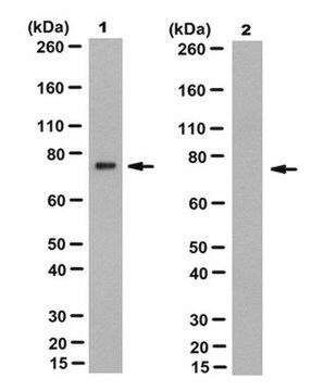
Immunoprecipitation Analysis: A representative immunoprecipitated Lamin A/C in culture supernatant (Data courtesy of Marie Lang, M.D., Stefan Schuchner, Ph.D. and Egon Ogris, M.D., Medical University of Vienna, Austria).


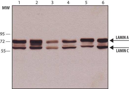
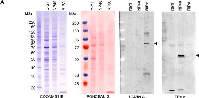
Western Blotting Analysis: A representative lot detected Lamin A/C in culture supernatants of various cell lines (Data courtesy of Marie Lang, M.D., Stefan Schuchner, Ph.D. and Egon Ogris, M.D., Medical University of Vienna, Austria).


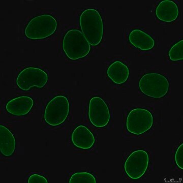
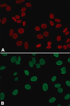
Immunocytochemistry Analysis: A 1:1,000 dilution from a representative lot detected Lamin A/C in HeLa cells.


Immunofluorescence Analysis: A representative lot detected Lamin A/C in the nuclear interior of HeLa cells (Data courtesy of Marie Lang, M.D., Stefan Schuchner, Ph.D. and Egon Ogris, M.D., Medical University of Vienna, Austria).

Qualität

Evaluated by Western Blotting in HeLa cell lysate.

Western Blotting Analysis: 0.2 µg/mL of this antibody detected Lamin A/C in HeLa cell lysate.

Zielbeschreibung

74 and 65 kDa observed. 74.14 kDa and 65.14 kDa for Lamin A and C. Uncharacterized bands may be observed in some lysate(s).

Physikalische Form

Protein G purified
Format: Purified
Purified mouse monoclonal antibody IgG2b in buffer containing 0.1 M Tris-Glycine (pH 7.4), 150 mM NaCl with 0.05% sodium azide.

Lagerung und Haltbarkeit

Stable for 1 year at 2-8°C from date of receipt.

Sonstige Hinweise

Concentration: Please refer to lot specific datasheet.

Haftungsausschluss

Unless otherwise stated in our catalog or other company documentation accompanying the product(s), our products are intended for research use only and are not to be used for any other purpose, which includes but is not limited to, unauthorized commercial uses, in vitro diagnostic uses, ex vivo or in vivo therapeutic uses or any type of consumption or application to humans or animals.

Not finding the right product?  

Try our Produkt-Auswahlhilfe.


Analysenzertifikate (COA)

Suchen Sie nach Analysenzertifikate (COA), indem Sie die Lot-/Chargennummer des Produkts eingeben. Lot- und Chargennummern sind auf dem Produktetikett hinter den Wörtern ‘Lot’ oder ‘Batch’ (Lot oder Charge) zu finden.

Besitzen Sie dieses Produkt bereits?

In der Dokumentenbibliothek finden Sie die Dokumentation zu den Produkten, die Sie kürzlich erworben haben.

Die Dokumentenbibliothek aufrufen

Unser Team von Wissenschaftlern verfügt über Erfahrung in allen Forschungsbereichen einschließlich Life Science, Materialwissenschaften, chemischer Synthese, Chromatographie, Analytik und vielen mehr..

Setzen Sie sich mit dem technischen Dienst in Verbindung.