Cell and Organelle Labeling with Fluorescent Antibodies
Prestige Antibodies® Powered by Atlas Antibodies for Organelle Labeling
The Human Protein Atlas Program has carefully selected three different human cell lines, A-431 epidermoid carcinoma, U-251 MG glioblastoma and U-205 osteosarcoma, for organelle mapping of the proteome. As Prestige Antibodies are studied by immunofluorescence (IF) staining, three well-characterized organelle markers for nuclei, microtubules and endoplasmic reticulum are used correspondingly as specific probes. The high-resolution confocal images are annotated based on sub-cellular localization. In addition, staining intensity and characteristics are also part of an overall assessment as well as comparison to literature (if available). All these factors contribute to a final reliability score publicly available on line. Among the Prestige Antibodies tested in immunofluorescence (IF), a set of specific antibodies is classified as organelle markers. These probes have been subjected to the well-established quality control by the HPA program, and are thus very well suited for sub-cellular localization studies by immunofluorescence (IF).
Cell Labeling Kits
PKH and CellVue® Fluorescent Cell Linker Kits provide fluorescent labeling of live cells over an extended period of time, with no apparent toxic effects. Fluorescent linker kits are effective for a variety of cell types and exhibit no significant leaking, or transfer from cell to cell. They provide stable, clear, intense, accurate and reproducible fluorescent labeling of cells. Patented cell linker technology incorporates aliphatic reporter molecules into the cell membrane lipid bilayer by selective partitioning.

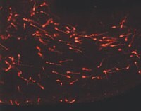
Figure 1.PKH26-labeled young migrating neurons invading the developing cortex. (Photo courtesy of H. Wichterle and Dr. A. Alvarez-Buylla, The Rockefeller University).
Four fluorescent linkers are available. PKH67 is a green fluorochrome (Product No. PKH67GL) with excitation (490 nm) and emission (504 nm) similar to fluorescein, and PKH26 is a red fluorochrome (Product No. PKH26GL) with excitation (551 nm) and emission (567 nm) characteristics compatible with rhodamine or phycoerythrin detection systems. PKH26 may also be excited by the 488 nm emission of an argon-ion laser. CellVue® Claret is a far-red fluorochrome that is independent of pH in physiologic ranges and has excitation (655 nm) and emission (675 nm) characteristics compatible with a red-diode laser. The linkers are physiologically stable and show minimal to no toxic side-effects on cell systems. Labeled cells retain both biological and proliferative activity and are ideal for cell tracking and cell-cell interaction studies.
The non-specific labeling mechanism of the cell linkers offers successful labeling of a wide variety of cell types. The linkers have been applied to both animal and plant cells as well as other membrane containing particles. The pattern of staining is dependent upon the cell type being labeled and the membrane of the cells. Although most applications center around general labeling (GL) methods involving membrane incorporation of the probes, the linkers may also be used for selective phagocytic cell labeling (PCL). Appearance of labeled cells may vary from bright "immunofluorescence" labeling to a punctate or patchy appearance. Since the labeling is not a saturation reaction, but rather a function of both dye and cell concentration, it is essential that the amount of dye available for incorporation be limited. Overlabeling of the cells will result in loss of membrane integrity and cell recovery.
Para seguir leyendo, inicie sesión o cree una cuenta.
¿No tiene una cuenta?