Atto Dyes and Tracy Dyes for Fluorescent Protein Labeling
We offer protein labeling kits based on two types of fluorescent dyes, the Atto dyes and the Tracy dyes (Figure 1). Both series of kits provide an easy and reliable way to fluorescently label purified proteins, enzymes, and antibodies (see procedure in Figure 2).

Figure 1.Protein fluorescent labeling kits available in our catalog.

Figure 2.Illustration of fluorescent labeling of proteins using Atto dyes or Tracy dyes.
Atto Dyes
The Atto dye Protein Labeling Kits are based on high quality Atto dyes that produce extraordinary bright fluorescent signals with a narrow fluorescence emission spectrum. Atto dyes efficiently label proteins, and the resulting conjugates demonstrate high relative fluorescence intensity when compared to other commercially available fluorescence dyes. Six protein labeling kits are available, each containing a reactive Atto dye specific for a commonly used excitation wavelength.
- Atto 488 is a superior alternative to fluorescein, producing conjugates with more photostability and brighter fluorescence. It can be excited with the same light sources as fluorescein dyes or Alexa® 488, and the same optical filter sets and instrument settings can be used to record the emission.
- For longer wavelengths, Atto 550 is efficiently excited by HeNe lasers and can be used as an alternative to rhodamine dyes, Cy3 or Alexa® 550, offering more intense brightness and increased photostability.
- Atto 594 is excited by HeNe lasers and can be used as alternative to Alexa® 594 or Texas Red®.
- Atto 647N, an extraordinary highly fluorescent dye, Atto 655, and Atto 633 are all well suited for excitation with HeNe or krypton lasers, similar to Cy5, Alexa® 647, Alexa® 633, and Alexa® 655.
The data in Tables 1 and 2 demonstrate the superior fluorescence of Atto dye-labeled proteins. Monoclonal anti-mouse IgG was labeled using Atto 550, Alexa® 555, or Cy3 dyes (Table 1). The relative fluorescence intensity signals (RFI) of the dye-labeled antibody solutions were measured using a spectrofluorimeter and a HeNe laser with an excitation wavelength of 544 nm; emission wavelength was optimized for each dye conjugate tested. The RFI signal of the Atto dye-labeled anti-mouse IgG at the emission wavelength of 570–585 nm was 2 to 4-fold higher than the signals from the other dye-antibody conjugates tested.
In a second experiment, monoclonal anti-mouse IgG was labeled using Atto 647N, Alexa® 647, or Cy5 (Table 2). The RFI signals of the dye-labeled antibody solutions were detected using a spectrofluorimeter and a HeNe laser with an excitation wavelength of 633 nm; emission wavelength was optimized for each dye conjugate tested. The RFI signal of the Atto dye-labeled anti-IgG at the emission wavelength of 650–665 nm was 6 to 13-fold higher than the signals from the other dye-antibody conjugates tested.
Tracy Dyes
Tracy 652 and Tracy 645, and Tracy 690 are three new near infrared (NIR) fluorescent dyes that are:
- Able to retain chemical stability of the activated N-hydroxysuccimide (NHS) ester under protein labeling conditions (basic pH, which prevents decomposition or hydrolysis that could result in poor labeling efficiency).
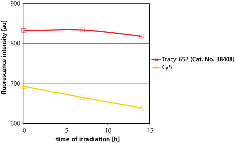
- Photostable with bright fluorescence intensity (Figure 3).
- Soluble in aqueous labeling media as the activated NHS ester, reducing precipitation that leads to inefficient protein labeling.
- Flexible for use in typical protein labeling protocols.
Tracy 652 and Tracy 645 surpass conventional fluorescent labels, such as Cy 5, in all of the above criteria. Other, more modern labels of the corresponding type are also deficient in one or more of the above criteria. Tracy 690 represents a high quality substitute for Cy 5.5.

Figure 3. Comparison of Photostabilities of Tracy 652 and Cy5.A 1 mM solution in methanol of each dye was irradiated in a white Pyrex flask by a 375 W white light lamp. The distance from lamp to probe was 20 cm and the probe maintained at room temperature. Fluorescence intensity (as au) was measured at λex,max and λem,max for each dye.
Protein Labeling Kits
All Atto Dye and Tracy Dye Protein Labeling Kits include:
- Five vials containing reactive fluorescent dye (optimized for 1 mg protein)
- Sodium bicarbonate reaction buffer solution, pH 9.5
- Phosphate buffer solution, pH 7.5
- Protein purification set containing 5 columns, 5 washing tubes and 5 sample collection tubes
- Technical bulletin with labeling protocol
Each kit contains sufficient amounts of reactive dye, buffers, and protein purification sets for performing five labeling reactions (1 mg protein each) and for the subsequent purification of the labeled proteins.
Para seguir leyendo, inicie sesión o cree una cuenta.
¿No tiene una cuenta?