Saltar al contenido
Merck
HomeProtein ExpressionEpidermal Growth Factor Receptor (EGFR) Signaling

Epidermal Growth Factor Receptor (EGFR) Signaling

Background
Epidermal Growth Factor Receptors (EGFR) and Ligands
Receptor Activation
EGFR Signaling
Ras/Raf Signaling Cascade
Phosphatidylinositol 3-kinase/Akt Signaling Cascade
Signal Transducers and Activators of Transcription (STAT) Pathway
Phospholipase Cγ Signaling
Nck/PAK Signaling Cascade
Cbl Mediated Endocytosis
EGFR Translocation into Nucleus
EGFRs in Development & Disease
Conclusion

Background

Epidermal growth factor receptor (EGFR) is the first discovered and prototypical member of receptor tyrosine kinase family (RTK) of receptors. It is activated by various ligands in extracellular milieu and transmits the cellular response to mediate various cellular activities, including cell proliferation, cell survival, growth, and development. EGFR is expressed in many organs and aberrant expression is implicated in various cancers. This review highlights various signaling components of EGFR pathway and their signal transduction pathways.

Epidermal Growth Factor Receptors (EGFR) and Ligands

The ErbB family comprises of four receptors, which include EGFR (ErbB-1/HER1), ErbB-2 (Neu, HER2), ErbB-3 (HER3), and ErbB-4 (HER4). This receptor tyrosine kinase family (RTK) of proteins has an extracellular ligand-binding domain, hydrophobic transmembrane domain, and a cytoplasmic tyrosine kinase domain (Table 1). ErbB receptors are activated by growth factors of EGF-family characterized by three disulphide-bonds that confer binding specificity. Additional structural motifs include immunoglobulin-like domains, heparin-binding sites and glycosylation sites1,2.

Table 1EGFR Receptors and Ligands

Receptor Activation

Receptor activation is triggered by the cascade of events on cell membrane.

  • Ligand binding: Each ligand binds to the extracellular domain of cognate ErbB receptors
  • Receptor dimerization: Ligand binding induces the formation of receptor homo or hetero dimers3.
  • Activation of kinase domain: Autophosphorylation of key tyrosine residues within carboxy terminal tail activates the receptor and acts as docking sites for proteins with Src homology 2 (SH2) and phosphotyrosine binding domain (PTB) to trigger cellular signaling3,4.
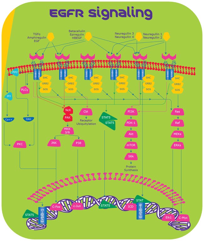
egfr signalling

Figure 1.EGFR Signaling

EGFR Signaling

Ras/Raf Signaling Cascade

Receptors: Dimerization of any two ErbB receptors

Key Functions: Cell survival and cell proliferation5

After receptor activation, the complex formed by Grb2 and Sos binds directly or through association of adapter protein Shc, to specific tyrosine residues on the receptor6,7. This leads to conformational change in Sos, which can recruit and activate Ras-GDP. Ras-GDP activates Raf-1, which further activates extracellular regulated kinases 1 and 2 (ERK1 and ERK2) mediated through mitogen-activated protein kinases (MAPK)8,9. Activated kinases eventually move into the nucleus to phosphorylate specific transcription factors like Elk1 and C-myc to induce cell proliferation.

Phosphatidylinositol 3-kinase/Akt Signaling Cascade

Receptors: Dimerization of ErbB2 with either ErbB4 or ErbB3

Key Functions: Cell growth, apoptosis resistance, cell invasion and migration10

Phosphatidylinositol comprises of p85 and p110 sub units that dock to the ErbB receptor to generate secondary messenger phosphatidylinositol 3,4,5-triphosphate, that further activates serine/threonine kinase AKT. Upon activation, AKT phosphorylates mTOR and subsequently S6K which mediates protein synthesis10.

Signal Transducers and Activators of Transcription (STAT) Pathway

Receptors: ErbB

Key Functions: Tumor progression, oncogenesis and angiogenesis11

STAT proteins docks to the phosphotyrosine residues of ErbB receptors via Src homology 2 domains and up on dimerization, translocate into the nucleus to promote the expression of specific target genes like Myc, Nos2, p21 and cytokines12.

Phospholipase Cγ Signaling

Receptors: ErbB1

Key Functions: Regulation of ion channels, cell migration, calcium-mediated signaling13

Phospholipase Cγ interacts with ErbB1 and hydrolyses phosphatidylinositol 4,5-diphosphate (PIP2) to generate inositol 1,3,5-triphosphate (IP3) and 1,2 diacylglycerol (DAG). IP3 increases intracellular calcium levels and DAG mediates activation of protein kinase C (PKC)13. Activated PKC in turn activates MAPK and c-Jun NH2-terminal kinase14.

Nck/PAK Signaling Cascade

Receptors: ErbB1

Key Functions: Cell survival and cell migration15

Nck is an adaptor protein containing SH2 domain that binds to EGF receptor and triggers downstream signaling. Nck activates PAK1 (p21/CDC42/Rac1-Activated Kinase-1) binding through SH3 domain. Activated PAK1 in turn activates JNKs (c-Jun Kinases) mediated by MEKK1 (MAP/ERK Kinase Kinase-1) and MKK4/7 (MAP Kinase Kinase-4/7), respectively. JNK translocates to the nucleus and phosphorylates transcription factors like c-Fos and c-Jun16.

Cbl Mediated Endocytosis

Receptors: ErbB1

Key Functions: Endocytosis

Following ligand binding, Cbl is a substrate that binds to the EGF receptors through SH2 domains or through GRB2 adaptor protein and triggers lysosomal degradation of receptors17.

EGFR Translocation into Nucleus

EGF Receptors have an ability to escape lysosomal degradation and translocate into the nucleus to mediate biological functions. In the nucleus, these receptors promote transcription of cell survival genes like Cyclin D1 gene and also act as cofactors for STAT and E2F1 transcription factors18. Nuclear localization of EGFR is implicated in disease severity by conferring resistance to growth inhibitory effects of anti-cancer mAbs19.

Our most popular, high-quality EGF proteins for your research use

EGFRs in Development and Disease

EGF receptors and their ligands are important for development of various organs; EGFR knockout models show embryonic lethality and defective tissues/organs. However, mutations in ErbB ligands do not show any lethal phenotype, because the complimentary pathways of other ligands compensate the abrogated signals of mutated ErbB ligand.20-25 EGFR are abnormally activated or expressed in many diseases. Specifically, their role in cancer progression is well investigated (Table 2).

Table 2Genetic alterations of EGFR receptors in human disorders

Conclusion

EGFR signaling has pleiotropic functions in development and regulates various physiological functions. The aberrant expression of EGF receptors has been increasingly implicated in various disorders. More research is yet to clearly uncover the role of EGFR signaling in development of various organs, including brain, heart, skin, kidneys, mammary glands, and lungs. Efforts to identify novel drugs with high efficacy, sustained drug activity and less cross reactivity to combat drug-resistant cancer conditions continue to pique lot of research interest.

1.
Arteaga C, Engelman J. 2014. ERBB Receptors: From Oncogene Discovery to Basic Science to Mechanism-Based Cancer Therapeutics. Cancer Cell. 25(3):282-303. https://doi.org/10.1016/j.ccr.2014.02.025
2.
Kovacs E, Zorn JA, Huang Y, Barros T, Kuriyan J. 2015. A Structural Perspective on the Regulation of the Epidermal Growth Factor Receptor. Annu. Rev. Biochem.. 84(1):739-764. https://doi.org/10.1146/annurev-biochem-060614-034402
3.
Capuani F, Conte A, Argenzio E, Marchetti L, Priami C, Polo S, Di Fiore PP, Sigismund S, Ciliberto A. 2015. Quantitative analysis reveals how EGFR activation and downregulation are coupled in normal but not in cancer cells. Nat Commun. 6(1): https://doi.org/10.1038/ncomms8999
4.
Zhang X, Gureasko J, Shen K, Cole PA, Kuriyan J. 2006. An Allosteric Mechanism for Activation of the Kinase Domain of Epidermal Growth Factor Receptor. Cell. 125(6):1137-1149. https://doi.org/10.1016/j.cell.2006.05.013
5.
Gaestel M. 2006. MAPKAP kinases ? MKs ? two's company, three's a crowd. Nat Rev Mol Cell Biol. 7(2):120-130. https://doi.org/10.1038/nrm1834
6.
Lowenstein E, Daly R, Batzer A, Li W, Margolis B, Lammers R, Ullrich A, Skolnik E, Bar-Sagi D, Schlessinger J. 1992. The SH2 and SH3 domain-containing protein GRB2 links receptor tyrosine kinases to ras signaling. Cell. 70(3):431-442. https://doi.org/10.1016/0092-8674(92)90167-b
7.
Batzer AG, Rotin D, Ureña JM, Skolnik EY, Schlessinger J. 1994. Hierarchy of binding sites for Grb2 and Shc on the epidermal growth factor receptor.. Mol. Cell. Biol.. 14(8):5192-5201. https://doi.org/10.1128/mcb.14.8.5192
8.
B H, S I R, J D. 1994. Interaction of Ras and Raf in intact mammalian cells upon extracellular stimulation. J Biol Chem. 269(6):3913-6.
9.
Liebmann C. 2001. Regulation of MAP kinase activity by peptide receptor signalling pathway: Paradigms of multiplicity. Cellular Signalling. 13(11):777-785. https://doi.org/10.1016/s0898-6568(01)00192-9
10.
Vivanco I, Sawyers CL. 2002. The phosphatidylinositol 3-Kinase?AKT pathway in human cancer. Nat Rev Cancer. 2(7):489-501. https://doi.org/10.1038/nrc839
11.
Niu G, Wright KL, Huang M, Song L, Haura E, Turkson J, Zhang S, Wang T, Sinibaldi D, Coppola D, et al. 2002. Constitutive Stat3 activity up-regulates VEGF expression and tumor angiogenesis. Oncogene. 21(13):2000-2008. https://doi.org/10.1038/sj.onc.1205260
12.
Bromberg J. 2002. Stat proteins and oncogenesis. J. Clin. Invest.. 109(9):1139-1142. https://doi.org/10.1172/jci0215617
13.
Patterson RL, van Rossum DB, Nikolaidis N, Gill DL, Snyder SH. 2005. Phospholipase C-?: diverse roles in receptor-mediated calcium signaling. Trends in Biochemical Sciences. 30(12):688-697. https://doi.org/10.1016/j.tibs.2005.10.005
14.
Scho?nwasser DC, Marais RM, Marshall CJ, Parker PJ. 1998. Activation of the Mitogen-Activated Protein Kinase/Extracellular Signal-Regulated Kinase Pathway by Conventional, Novel, and Atypical Protein Kinase C Isotypes. Mol. Cell. Biol.. 18(2):790-798. https://doi.org/10.1128/mcb.18.2.790
15.
Ye DZ, Field J. 2012. PAK signaling in cancer. Cellular Logistics. 2(2):105-116. https://doi.org/10.4161/cl.21882
16.
Tomar A, Schlaepfer DD. 2010. A PAK-Activated Linker for EGFR and FAK. Developmental Cell. 18(2):170-172. https://doi.org/10.1016/j.devcel.2010.01.013
17.
Yu P, Fan Y, Qu X, Zhang J, Song N, Liu J, Liu Y. 2016. Cbl-b regulates the sensitivity of cetuximab through ubiquitin-proteasome system in human gastric cancer cells. J Buon. 21(4):867-873.
18.
Lo H, Hsu S, Ali-Seyed M, Gunduz M, Xia W, Wei Y, Bartholomeusz G, Shih J, Hung M. 2005. Nuclear interaction of EGFR and STAT3 in the activation of the iNOS/NO pathway. Cancer Cell. 7(6):575-589. https://doi.org/10.1016/j.ccr.2005.05.007
19.
Lo H, Xia W, Wei Y, MA, Huang S, Hung M. 2005. Novel prognostic value of nuclear epidermal growth factor receptor in breast cancer. Cancer Res. 65(1):338-48.
20.
Miettinen PJ, Berger JE, Meneses J, Phung Y, Pedersen RA, Werb Z, Derynck R. 1995. Epithelial immaturity and multiorgan failure in mice lacking epidermal growth factor receptor. Nature. 376(6538):337-341. https://doi.org/10.1038/376337a0
21.
Sibilia M, Wagner E. 1995. Strain-dependent epithelial defects in mice lacking the EGF receptor. Science. 269(5221):234-238. https://doi.org/10.1126/science.7618085
22.
Park S, Miller R, Krane I, Vartanian T. 2001. The erbB2 gene is required for the development of terminally differentiated spinal cord oligodendrocytes. 154(6):1245-1258. https://doi.org/10.1083/jcb.200104025
23.
Leu M. 2003. Erbb2 regulates neuromuscular synapse formation and is essential for muscle spindle development. 130(11):2291-2301. https://doi.org/10.1242/dev.00447
24.
S L E, K S O, N G, L L, G F, M B, L H L, M W M. 1997. ErbB3 is required for normal cerebellar and cardiac development: a comparison with ErbB2-and heregulin-deficient mice. Development. 124(24):4999-5011.
25.
Golub MS, Germann SL, Lloyd K. 2004. Behavioral characteristics of a nervous system-specific erbB4 knock-out mouse. Behavioural Brain Research. 153(1):159-170. https://doi.org/10.1016/j.bbr.2003.11.010
26.
Maheswaran S, Sequist LV, Nagrath S, Ulkus L, Brannigan B, Collura CV, Inserra E, Diederichs S, Iafrate AJ, Bell DW, et al. 2008. Detection of Mutations inEGFRin Circulating Lung-Cancer Cells. N Engl J Med. 359(4):366-377. https://doi.org/10.1056/nejmoa0800668
27.
Montagut C, Dalmases A, Bellosillo B, Crespo M, Pairet S, Iglesias M, Salido M, Gallen M, Marsters S, Tsai SP, et al. 2012. Identification of a mutation in the extracellular domain of the Epidermal Growth Factor Receptor conferring cetuximab resistance in colorectal cancer. Nat Med. 18(2):221-223. https://doi.org/10.1038/nm.2609
28.
Rosell R, Moran T, Queralt C, Porta R, Cardenal F, Camps C, Majem M, Lopez-Vivanco G, Isla D, Provencio M, et al. 2009. Screening for Epidermal Growth Factor Receptor Mutations in Lung Cancer. N Engl J Med. 361(10):958-967. https://doi.org/10.1056/nejmoa0904554
29.
Liu Z, Hou P, Ji M, Guan H, Studeman K, Jensen K, Vasko V, El-Naggar AK, Xing M. 2008. Highly Prevalent Genetic Alterations in Receptor Tyrosine Kinases and Phosphatidylinositol 3-Kinase/Akt and Mitogen-Activated Protein Kinase Pathways in Anaplastic and Follicular Thyroid Cancers. The Journal of Clinical Endocrinology & Metabolism. 93(8):3106-3116. https://doi.org/10.1210/jc.2008-0273
30.
Gao SP, Mark KG, Leslie K, Pao W, Motoi N, Gerald WL, Travis WD, Bornmann W, Veach D, Clarkson B, et al. 2007. Mutations in the EGFR kinase domain mediate STAT3 activation via IL-6 production in human lung adenocarcinomas. J. Clin. Invest.. 117(12):3846-3856. https://doi.org/10.1172/jci31871
31.
Fukushige S, Murotsu T, Matsubara K. 1986. Chromosomal assignment of human genes for gastrin, thyrotropin (TSH)-? subunit and C-erbB-2 by chromosome sorting combined with velocity sedimentation and southern hybridization. Biochemical and Biophysical Research Communications. 134(2):477-483. https://doi.org/10.1016/s0006-291x(86)80445-4
32.
Slamon D, Godolphin W, Jones L, Holt J, Wong S, Keith D, Levin W, Stuart S, Udove J, Ullrich A, et al. 1989. Studies of the HER-2/neu proto-oncogene in human breast and ovarian cancer. Science. 244(4905):707-712. https://doi.org/10.1126/science.2470152
33.
Qiu Y, Ravi L, Kung H. 1998. Requirement of ErbB2 for signalling by interleukin-6 in prostate carcinoma cells. Nature. 393(6680):83-85. https://doi.org/10.1038/30012
34.
Harskamp LR, Gansevoort RT, van Goor H, Meijer E. 2016. The epidermal growth factor receptor pathway in chronic kidney diseases. Nat Rev Nephrol. 12(8):496-506. https://doi.org/10.1038/nrneph.2016.91
35.
Scafidi J, Hammond TR, Scafidi S, Ritter J, Jablonska B, Roncal M, Szigeti-Buck K, Coman D, Huang Y, McCarter RJ, et al. 2014. Intranasal epidermal growth factor treatment rescues neonatal brain injury. Nature. 506(7487):230-234. https://doi.org/10.1038/nature12880
36.
Campbell P, Morton PE, Takeichi T, Salam A, Roberts N, Proudfoot LE, Mellerio JE, Aminu K, Wellington C, Patil SN, et al. 2014. Epithelial Inflammation Resulting from an Inherited Loss-of-Function Mutation in EGFR. Journal of Investigative Dermatology. 134(10):2570-2578. https://doi.org/10.1038/jid.2014.164
Inicie sesión para continuar.

Para seguir leyendo, inicie sesión o cree una cuenta.

¿No tiene una cuenta?