Identification of Mycobacteria
Anandi Martin, PhD.
Institute of Tropical Medicine, Mycobacteriology Unit, Belgium
Microbiology Focus Edition 2.2

Figure 1. Typical colony of Mycobacterium tuberculosis seen under a microscope with 10x magnification
The diseases produced by species of the genus Mycobacterium are important causes of morbidity and mortality in the world. The identification of mycobacteria to the species level is important because of the clinical significance; some species are pathogenic while others are not.
Traditionally, mycobacteria are identified by phenotypic methods, based on culture, such as morphological characteristics, growth rates, preferred growth temperature, pigmentation and on a series of biochemical tests. Testing is laborious, difficult and time-consuming, requiring several weeks for adequate growth, and sometimes misidentification may occur because different species may have indistinguishable morphological and biochemical profiles. Different culture media are in use for the isolation of mycobacteria. The most common are based on eggs called “Löwenstein-Jensen medium” and contain high concentrations of malachite green to overcome contamination with other bacteria.
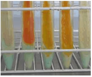
In the last decade, several commercial systems for mycobacterial culture based on liquid media have been introduced. Liquid culture media have been proven to be significantly more sensitive than egg-based solid media for the isolation of mycobacteria from clinical specimens. M. tuberculosis bacilli are slow-growing mycobacteria which means that in primary isolation they hardly show any visible growth during the first week of culture. On egg-based media they produce characteristic nonpigmented colonies, with a general rough and dry appearance simulating breadcrumbs. On agar based media, the colonies appear flat, dry and rough with irregular edges. M. tuberculosis is niacin positive, is inhibited by p-nitrobenzoic acid and display nitratase activity. Additional tests that confirm an isolate as M. tuberculosis are susceptible to pyrazinamide, growth on thiophene carboxylic acid hydrazide (TCH), absence of catalase production at 68 °C and absence of iron uptake.

Figure 2. Mycobacteria colonies on TB-Medium Base according to Löwenstein-Jensen
The Ziehl-Neelsen staining for the direct detection of mycobacteria by microscopy is used to identify acid fast bacilli. The lipid rich cell wall of mycobacteria makes it resistant to Gram stain. It can also be used to stain few other bacteria like Nocardia. The reagents used for the staining are carbolfuchsin, acid-alcohol and methylene blue. Acid fast bacilli appear bright red after staining.
Non-tuberculous mycobacteria (NTM) are ubiquitous organisms that are frequently isolated from environmental sources, including surface water, tap water, and soil. The NTM species most frequently associated with pulmonary disease are M. avium, M. kansasii and M. abscessus. Injury cutaneous/ subcutaneous infections have been attributed to rapidly growing mycobacteria. M. fortuitum, M. abscessus, and M. chelonae are thought to be caused by local environmental strains or contaminated commercial surgical materials, devices or solutions for injection. Rapidly growing mycobacteria often grow on classical bacterial culture media, especially on blood agar plates, however, due to their delay in forming visible colonies (up to 10 days), they are usually not detected in the routine bacteriology laboratory. They can also be isolated on most media available for the isolation of mycobacteria. Although the optimum temperature for most species is 30-32 °C, they also grow at 36-37 °C, the standard temperature for isolation of the M. tuberculosis.
In the last decade, advances in molecular methods have facilitated the rapid and reliable identification of many mycobacterial species. Nucleic acid probes, species-specific PCR, reverse hybridization and 16S rRNA sequencing have been evaluated for application in clinical laboratories. The first method commercially available was the AccuProbe (Gen- Probe Inc.), based on species-specific DNA probes that hybridize to rRNA for the identification of several important mycobacteria, including the M. tuberculosis complex, M. avium, M. intracellulare, the M. avium complex, M. kansasii and M. gordonae. More recently, other molecular commercial systems have also been introduced for the rapid identification of M. tuberculosis complex: the INNO- LiPA MYCOBACTERIA v2 (Innogenetics NV, Ghent, Belgium), and the Geno- Type MTBC and GenoType Mycobacterium (Hain Lifesciences, Nehren, Germany). INNO-LiPA MYCOBACTERIA v2 is a line probe assay that simultaneously detects and identifies the genus Mycobacterium and 16 different mycobacterial species. It is based on nucleotide differences in the 16S-23S rRNA gene spacers. The GenoType MTBC and GenoType Mycobacterium are also based on the reverse line probe hybridization assay and are intended for the differentiation of members of the M. tuberculosis complex and for the identification of 35 species of mycobacteria including M. tuberculosis, respectively. The GenoType MTBC is based on a 23S rRNA gene fragment specific for the M. tuberculosis complex, together with gyrB sequence polymorphisms, and the RD1 deletion for identification of M. bovis BCG. Several ‘in-house’ techniques are also available with sequencing of the 16S rRNA gene as the reference standard to which all other new techniques are generally compared.

Figure 3. Different mycobacteria species grown on TB-Medium Base according to Löwenstein- Jensen
References
Para seguir leyendo, inicie sesión o cree una cuenta.
¿No tiene una cuenta?