Saltar al contenido
Merck
Todas las fotos(1)

Key Documents

301M-1

Sigma-Aldrich

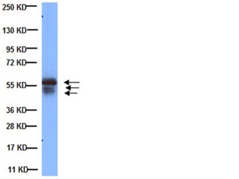
Cytokeratin, LMW (AE1) Mouse Monoclonal Antibody

Iniciar sesiónpara Ver la Fijación de precios por contrato y de la organización


About This Item

UNSPSC Code:
12352200
NACRES:
NA.41

biological source

mouse

Quality Level

100
500

conjugate

unconjugated

antibody form

culture supernatant

antibody product type

primary antibodies

clone

AE1, monoclonal

description

For In Vitro Diagnostic Use in Select Regions (See Chart)

form

buffered aqueous solution

species reactivity

human

packaging

vial of 0.1 mL concentrate (301M-14)
vial of 0.5 mL concentrate (301M-15)
bottle of 1.0 mL predilute (301M-17)
vial of 1.0 mL concentrate (301M-16)
bottle of 7.0 mL predilute (301M-18)

manufacturer/tradename

Cell Marque

technique(s)

immunohistochemistry (formalin-fixed, paraffin-embedded sections): 1:100-1:500

isotype

IgG1κ

control

prostate

shipped in

wet ice

storage temp.

2-8°C

visualization

cytoplasmic

Gene Information

human ... KRT1(3848)

Categorías relacionadas

General description

Anti-cytokeratin, low molecular weight (AE1) labels most acidic keratins; therefore, it is a broadly reactive antibody staining most epithelia and their neoplasms. Members of the acidic and basic cytokeratin subfamilies are found together in pairs; each epithelium contains at least one acidic and one basic keratin so this antibody can show the distribution of keratin containing cells in epithelia. This antibody has shown great sensitivity and broad specificity for keratins under various conditions of fixation and staining. Anti-low molecular weight cytokeratin (AE1) is particularly suited to distinguish poorly differentiated carcinomas from non-epithelial neoplasms. This marker stains both normal and neoplastic cells of epithelial origin.

Quality


IVD

IVD

IVD

RUO

Linkage

Cytokeratin, LMW Positive Control Slides, Product No. 301S, are available for immunohistochemistry (formalin-fixed, paraffin-embedded sections).

Physical form

Solution in Tris Buffer, pH 7.3-7.7, with 1% BSA and <0.1% Sodium Azide

Preparation Note

Download the IFU specific to your product lot and formatNote: This requires a keycode which can be found on your packaging or product label.

Other Notes

For Technical Service please contact: 800-665-7284 or email: service@cellmarque.com

Legal Information

Cell Marque is a trademark of Merck KGaA, Darmstadt, Germany

¿No encuentra el producto adecuado?  

Pruebe nuestro Herramienta de selección de productos.


Certificados de análisis (COA)

Busque Certificados de análisis (COA) introduciendo el número de lote del producto. Los números de lote se encuentran en la etiqueta del producto después de las palabras «Lot» o «Batch»

¿Ya tiene este producto?

Encuentre la documentación para los productos que ha comprado recientemente en la Biblioteca de documentos.

Visite la Librería de documentos

A R Judkins et al.
American journal of clinical pathology, 110(5), 641-646 (1998-11-05)
Immunohistochemistry occasionally is used to determine the lineage of entirely necrotic tumors. However, the sensitivity and specificity of antibodies on necrotic tissue are unknown. To determine the usefulness of immunohistochemistry with necrotic lesions, a series of 24 known tumors consisting
R Kitazawa et al.
Virchows Archiv : an international journal of pathology, 435(2), 137-142 (1999-12-22)
A 69-year-old man had a hepatic tumour occupying the left and half of the right lobe, with portal vein thrombus. There were hypercalcaemia and hypophosphataemia with increased nephrogenous cyclic adenosine monophosphate; bone metastases were excluded. Serum parathyroid hormone-related protein (PTHrP)
A J Demetris et al.
The American journal of pathology, 149(2), 439-448 (1996-08-01)
The ductular reaction to acute submassive necrosis was studied in human livers removed at the time of orthotopic liver transplantation. Single, double, and triple immunohistochemical labeling in combination with morphometry was used to analyze the phenotype and proliferative and apoptotic
Diagnostic Immunohistochemistry: Theranostic and Genomic Applications, 166-174 (2002)
V Eusebi et al.
The American journal of surgical pathology, 14(8), 737-747 (1990-08-01)
We present four cases of a malignant thyroid tumor showing morphologic, immunocytochemical, and ultrastructural features of endothelial cell differentiation. The tumor cells had epithelioid features and displayed strong immunoreactivity for keratin. There was no evidence of follicular or C-cell differentiation

Nuestro equipo de científicos tiene experiencia en todas las áreas de investigación: Ciencias de la vida, Ciencia de los materiales, Síntesis química, Cromatografía, Analítica y muchas otras.

Póngase en contacto con el Servicio técnico